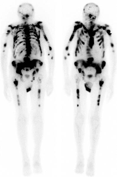
CASE 17 A 72-year-old man with a history of metastatic prostate cancer is referred for a bone scan because of pain in the left shoulder and lower neck (Fig. 17.1). Fig. 17.1 • A 20 mCi dose of 99mTc-MDP is administered intravenously. • Whole-body images of the skeleton are obtained 3 hours after tracer administration. • A 1024 × 256 matrix is used. • Emphasize the importance of oral hydration to improve soft tissue and bladder clearance. Whole-body images show intense tracer uptake throughout the axial skeleton and proximal portions of the appendicular skeleton. Little uptake is noted in the soft tissues, including the kidneys. A small amount of tracer is noted in the urinary bladder. • Disseminated prostate cancer. The patient had previously received 89Sr therapy with little relief of his pain. He was therefore scheduled for external beam therapy to the neck and left shoulder. A bone scan with disseminated, intense uptake is often called a superscan
Clinical Presentation
Technique
Image Interpretation
Diagnosis and Clinical Follow-Up
Discussion
![]()
Stay updated, free articles. Join our Telegram channel


Full access? Get Clinical Tree


