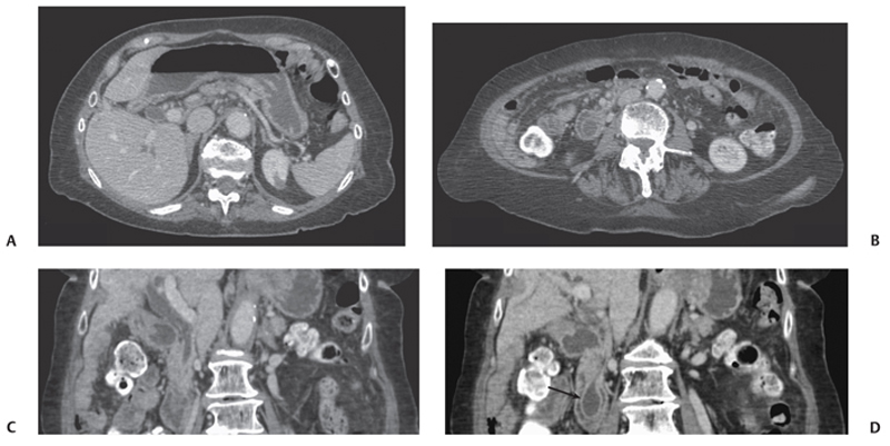
CASE 18 A 52-year-old Asian woman complains of vague abdominal pain. Fig. 18.1 (A–D) Axial and coronal contrast-enhanced CT images show mild prominence of the extra- and intrahepatic bile duct. The dilated duct leads to a hypodense dilated distal choledochocele within the duodenum (arrow). Axial and coronal contrast-enhanced computed tomography (CT) image shows mild prominence of the extra- and intrahepatic bile duct. The dilated duct leads to a hypodense dilated distal end within the duodenum (Fig. 18.1). Choledochocele (type III choledochal cyst; Table 18.1)
Clinical Presentation
Radiologic Findings
Diagnosis
| Type | Description | Occurrence (%) |
|---|---|---|
| I | Solitary extrahepatic cyst | 50 to 70 |
| II | Extrahepatic supraduodenal diverticulum | 1 to 3 |
| III | Intraduodenal diverticulum (choledochocele) | 1 to 4 |
| IV | Extrahepatic and intrahepatic dilatation | 15 to 33 |
| V | Multiple intrahepatic cysts (Caroli disease) | < 1 |
Related posts:


Stay updated, free articles. Join our Telegram channel

Full access? Get Clinical Tree


