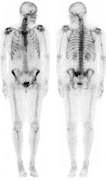
CASE 18 A 50-year-old woman presents with a history of breast cancer (Fig. 18.1). Fig. 18.1 • A 20 mCi dose of 99mTc-MDP is administered intravenously. • Whole-body or spot images of the skeleton are obtained 3 hours after tracer administration. • Emphasize the importance of oral hydration to improve soft tissue and bladder clearance. Whole-body views (Fig. 18.1) demonstrate tracer uptake in the upper cervical spine, midshaft of the right humerus, and left sacroiliac joint, and a smaller focus in the adjacent sacrum. Also noted is uptake in the soft tissues of the right upper quadrant of the abdomen.
Clinical Presentation
Technique
Image Interpretation
Related posts:


Stay updated, free articles. Join our Telegram channel

Full access? Get Clinical Tree


