18 Mega Cisterna Magna
18.1 Case Presentation
18.1.1 History
A 15-year-old female patient presents with a history of a one-time seizure episode.
18.2 Imaging Analysis
18.2.1 Imaging Findings and Impression
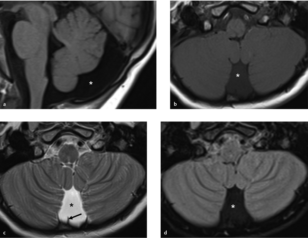
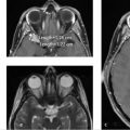
Brain MR images sagittal T1 (▶ Fig. 18.1a), axial T1 (▶ Fig. 18.1b), axial T2 (▶ Fig. 18.1c), and axial T2 fluid-attenuated inversion recovery (FLAIR; (▶ Fig. 18.1d) images are displayed. There is widening of the subarachnoid spaces posterior and inferior to the vermis (asterisk), without any mass effect or displacement of the falx cerebelli (black arrow). The cerebellar vermis and fourth ventricle are normal. Findings are consistent with a mega cisterna magna (MCM).
18.3 Differential Diagnosis
MCM:
It is a normal variant and corresponds to a focal enlargement of subarachnoid space in the posterior and inferior midline posterior fossa.
Posterior fossa arachnoid cyst:
It is an extra-axial collection of cerebrospinal fluid (CSF) delineated by arachnoid that does not directly communicate with the ventricular system or the subarachnoid space.
Retrocerebellar arachnoid cyst pushes the vermis forward, whereas an MCM pushes the vermis up from foramen magnum. 1 , 2 , 3
Midline arachnoid cyst demonstrates mass effect and deviation of the falx cerebelli.
Dandy–Walker malformation and variants:
Cystic dilatation of the fourth ventricle, enlarged posterior fossa, small hypoplastic vermis superiorly rotated, and elevated torcula are some symptoms.
In MCM, there is no malformation of vermis, cerebellum, or other posterior fossa components. 3 , 4 , 5
Blake’s pouch cyst:
Persistent Blake’s pouch cysts occur due to a failed perforation of the foramen of Magendie.
Findings include an infravermian cyst that communicates with the fourth ventricle without communication with the cisterna magna posteriorly.
It is often associated with mild upward displacement and rotation of vermis but normally formed vermis, normally positioned torcula.
Choroid plexus can extend from the fourth ventricle into the superior portion of the cyst.
Related posts:
Stay updated, free articles. Join our Telegram channel

Full access? Get Clinical Tree


