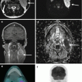
Fig. 6.1
FDG PET/CT of a 34-year-old patient with suspected relapse in previously treated Hodgkin disease showed intensely FDG-avid mediastinal disease (big arrow). Intense physiological brown fat uptake in bilateral neck/supraclavicular fossae (small arrows) can hamper the assessment of FDG uptake in the small nodes in these sites

Fig. 6.2
Pretreatment FDG PET/CT of a 40-year-old patient with Hodgkin disease showed intensely FDG-avid bulky cervical lymphadenopathy (long arrow) extending into superior mediastinum (stage II). Linear intense endometrial uptake (short arrow) was confidently reported as physiological uptake because of patient’s menstrual history in the scan preparation checklist
A common physiological process which can affect the lymphoma FDG PET/CT quality is brown fat uptake. It is common in colder environment and in children but can also occur in adults. Neck/supraclavicular, mediastinal, axillary and paraspinal regions are common sites masking the view. The symmetrical uptake in typical locations together with reassuring CT findings will exclude pathology. Brown fat uptake may be decreased by keeping a warm environment at FDG uptake phase. Beta-blocker and diazepam can also be used in advance.
Physiological uptake in muscles can sometimes be confused with nodal uptake if focal and close to nodal sites, e.g. scalene.
Some of the false-positive benign FDG-avid conditions include reactive nodes; inflammation related to injection, immunisation, trauma, biopsy, surgery, chemotherapy or radiotherapy; granulomatous inflammation; and acute and chronic infection such as TB, fungal infection, toxoplasmosis, etc. The recommended time between treatments and FDG PET/CT is at least 10 days for chemotherapy (6–8 weeks post chemotherapy if end of treatment scans), 2 weeks for growth factors (G-CSF and GM-CSF), 6 weeks for surgery and 3 months for radiotherapy (Figs. 6.3 and 6.4).


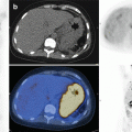
Fig. 6.3
Baseline FDG PET/CT of a lymphoma patient showing a reactive FDG-avid focus at the recently inserted Hickman line site (images from left to right – Non attenuation corrected PET, Attenuation corrected PET, Fused PET/CT)

Fig. 6.4
FDG PET/CT of a lymphoma patient showing an FDG-avid small reactive left axillary node. Patient gave a history of flu vaccination in the left arm a week before coming to PET/CT
Lymphoma PET/CT assessment now widely utilises Deauville criteria. Persistent or new lesions (Deauville score 4 or 5) represent positive scans, but false-positive results could lead to incorrect treatment decisions. The new FDG-avid areas which are unlikely to be lymphoma are marked as “X” in Deauville criteria. New or residual uptake while other known disease locations respond may indicate a separate pathology such as inflammation/infection or another tumour. It is important to correlate clinical, laboratory and imaging results in multidisciplinary settings when unexpected results or mixed response arise. Residual PET positve lesions may need pathology confirmation (e.g. when salvage treatment is considered) or follow up imaging (e.g. when clinical probability of active disease is low).
6.2.2 False Negative
FDG PET/CT can be false negative in small lesions, in lesions masked by adjacent high physiological uptake structures, and in some indolent lymphoma. The PET/CT response relies on treatment-induced changes in FDG uptake which are difficult to detect if the disease is not FDG avid or too small. Common types of lymphoma, e.g. HD and NHL such as DLCBL, are usually highly FDG avid. Some lymphoma such as MALT lymphoma and small lymphocytic lymphoma have low FDG avidity. Lymphoma involving the skin and mucosa may be less FDG avid or difficult to detect. In non-FDG-avid lymphoma, response assessment should be by standard CT instead of FDG PET.
6.3 Anatomy Based
6.3.1 Lymphatic Sites
Waldeyer’s ring
Waldeyer’s ring is often involved in NHL, but these lymphoid tissues frequently have high physiological/reactive FDG uptake. Their interpretation can be difficult when FDG uptake is asymmetrical. Clinical correlation (infection, tonsillectomy, etc.) and review of CT or MRI are crucial to check any mass lesion which would require direct examination and histology confirmation (Fig. 6.5).


Fig. 6.5
Baseline FDG PET/CT of a patient with nodal lymphoma showed intensely FDG-avid mediastinal and left neck nodal disease. There was asymmetrical increased left tonsillar uptake without CT abnormality. Physiological/reactive uptake is usually symmetrical but can also be asymmetrical
Lymph Nodes
Although lymphoma can affect any nodal sites, FDG PET false-positive reactive/inflammatory nodes are relatively common in neck, axillary and inguinal regions. Reactive nodes may have benign CT morphology (small, bean shaped, with fatty hilum, without necrosis or extracapsular spread, etc.). Assessment of drainage areas may also point to a possible aetiology, e.g. FDG extravasation, immunisation, surgical procedures, inflammation/infection, etc.
Granulomatous inflammation such as sarcoid reactions can appear following chemotherapy and can be misinterpreted as lymphoma. Typical pattern of sarcoid is FDG-avid bilateral hilar and mediastinal lymphadenopathy but can also be generalised lymphadenopathy. Acute as well as chronic infection such as TB, toxoplasma and fungal infection can also result in FDG-avid lymphadenopathy (Figs. 6.6 and 6.7).


Fig. 6.6
A 49-year-old man with intravascular large B cell NHL of pelvic and mediastinal disease. (Upper row) End-of-treatment FDG PET/CT showing intensely FDG-avid bilateral hilar and mediastinal lymphadenopathy. Clinical and imaging results were inconclusive for residual disease or sarcoid reaction. Mediastinal nodal biopsy confirmed sarcoidosis (false-positive inflammatory FDG uptake). (Lower row) FDG PET/CT 2-year post-treatment showed widespread focal FDG-avid bone marrow and soft tissue relapses including cutaneous and intramuscular deposits but no active disease in the mediastinum (lymphoma can relapse following complete metabolic response)

Fig. 6.7
FDG PET/CT of a 28 year old lady with suspected relapse Hodgkin Disease showedintensely FDG avid left neck and bilateral axillary lymphadenopathy. Excisional biopsy of the left axillary node showed no evidence of lymphoma but showed toxoplasma infection. (False positive infection FDG uptake)
Other potential false-positive inflammatory conditions include sclerosing mesenteritis (panniculitis) which can appear as FDG-avid diffuse ‘hazy mesentery’ or focal nodularity/masses and can mimic active mesenteric nodal lymphoma. Development of new mesenteric/peritoneal lesions while other sites show FDG response may indicate panniculitis rather than new lymphoma (Fig. 6.8). Similarly inflammatory conditions such as fibrosing mediastinitis can mimick mediastnal lymphoma.


