7.2 Case 2: Early Stage Unfavourable Disease
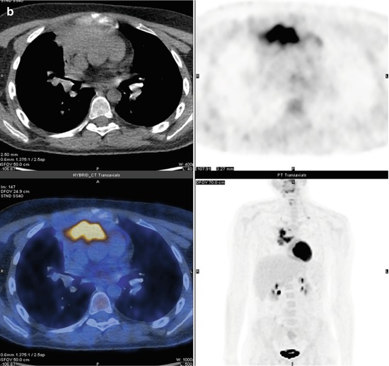
Baseline staging scan in 32-year-old female patient with Hodgkin’s lymphoma. PET/CT scan shows early stage (stage II) unfavourable disease. There is bulky disease within the mediastinum, involvement of three nodal sites (right cervical, left cervical, mediastinum) and suspicion of contiguous involvement of left lung from a nodal site.
7.2.1 Teaching Points
Hodgkin’s lymphoma is divided into early (stage I or II) and advanced (stage III or IV) disease. Early disease is further subdivided into favourable and unfavourable disease based on combination of imaging, laboratory and clinical markers and has different treatments. Three imaging markers of unfavourable disease include disease bulk, involvement of three or more nodal sites and extranodal (E) involvement.

7.3 Case 3: Splenic Involvement on PET/CT
Baseline Staging PET/CT in 22-year-old female with Hodgkin’s lymphoma. PET/CT shows stage IV disease. There is also abnormal increased heterogeneous FDG uptake within the spleen with reversal of normal hepato-splenic ratio.
7.3.1 Teaching Points
Normally in untreated patients, FDG activity within the spleen is less than the liver.
Splenic involvement on PET can be seen as diffuse increased uptake with reversal of normal hepato-splenic ratio, heterogeneous increased splenic activity or focal/multifocal avid splenic lesions.

7.4 Case 4: Reactive Marrow Activity Pre-Treatment
Staging PET scan in 21-year-old male with newly diagnosed Hodgkin’s lymphoma. Note the prominent diffuse FDG activity within bone marrow.
7.4.1 Teaching Points
Diffuse marrow activity in newly diagnosed HL is a relatively common finding and does not indicate bone marrow involvement being felt to represent reactive bone marrow activity.
7.5 Case 5: Bone Marrow Involvement
Baseline staging PET/CT in 22-year-old female with Hodgkin’s lymphoma. PET/CT shows stage IV disease with multifocal uptake within bone marrow. Two sites are shown on axial CT and fused PET/CT images in left humerus and sacrum.
7.5.1 Teaching Points
Multifocal FDG-avid lesions are typical patterns of bone marrow involvement in HL.
Often, as in this case, CT images show no morphological abnormality.
PET/CT is the most sensitive staging modality for assessment of bone marrow involvement and is superior to CT and routine bone marrow biopsy.

7.6 Case 6: Brown Fat Activity
Baseline staging scan in 20-year-old female with Hodgkin’s lymphoma. There is FDG-avid nodal disease within the mediastinum but also florid FDG activity within fat planes of the neck and shoulder girdle.
7.6.1 Teaching Points
FDG activity in brown fat is a common physiological variant. This can usually be distinguished from nodal activity with PET/CT but in florid cases can adversely impact the assessment of scans.
Techniques to minimise brown fat activity include keeping patient warm and giving diazepam or propranolol.

7.7 Case 7: Thymic Hyperplasia
PET/CT scans at baseline (A), end of treatment (B) and 7 months post end of treatment (C) in a 21-year-old female patient. End of treatment scan shows diffuse FDG activity in anterior mediastinum in soft tissue which has a thymic configuration. This was taken as representing thymic hyperplasia rather than disease despite the anterior mediastinum clearly being an involved disease site on the baseline scan. The patient was monitored clinically with a follow-up scan (C) showing persistent thymic activity. She remained well with no further treatment.
7.7.1 Teaching Points
Thymic hyperplasia is a common finding post chemotherapy.

7.8 Case 8: Reactive Bone Marrow and Spleen Activation
PET/CT scan post chemotherapy in 42-year-old male patient with lymphoma. There is diffuse increased FDG activity within the spleen and bone marrow. Neither site was involved at baseline. The findings are typical for post chemotherapy bone marrow and splenic activation.
7.8.1 Teaching Points
Reactive bone marrow and splenic activation occurs commonly post chemotherapy and GCF administration and should not be called active disease.
7.9 Case 9: Metabolic Remission
Interim (post two cycles of chemotherapy) PET/CT in a 22-year-old female patient with Hodgkin’s lymphoma. There are mildly enlarged residual nodes in the mediastinum which show very low-grade FDG activity. The uptake score is 2 (not greater than mediastinum background).
7.9.1 Teaching Points
An uptake score of 2 is considered to represent a negative scan on both interim and end of treatment scans.
Mediastinal activity should be assessed in large vessels, taking care to avoid uptake along walls of vessel.

7.10 Case 10: Interim PET Scan with Uptake Score of 3
Interim PET/CT post two cycles of ABVD chemotherapy in 21-year-old male patient with Hodgkin’s lymphoma. There is low-grade FDG activity within soft tissue in the anterior mediastinum. The uptake score is 3 (greater than the mediastinum but not greater than the liver).
7.10.1 Teaching Points
Interim PET is predictive of outcome in Hodgkin’s lymphoma.
A score of 3 is considered adequate response to chemotherapy if patient is to receive standard treatment.
If treatment is intended to be de-escalated as a result of a negative PET scan, then a lower threshold of 2 for a negative PET scan may be appropriate.

7.11 Case 11: Interim PET Scan with Uptake Score of 4
Baseline and interim (post cycles ABVD chemotherapy) scans in a 24-year-old female with advanced Hodgkin’s lymphoma. The highest uptake at disease site on interim scan is in an infraclavicular node (arrows) with an uptake score of 4 (moderately greater than liver).
7.11.1 Teaching Points
A score of 4 on an interim PET scan is considered positive for active disease.

7.12 Case 12: Interim PET Scan with Uptake Score of 5
Baseline (A) and interim (B, post two cycles ABVD chemotherapy) PET/CT scans in 20-year-old female. There is intense residual FDG uptake on interim PET scan with an uptake score of 5 (uptake markedly above liver).
7.12.1 Teaching Points
A score of 5 on an interim PET scan represents inadequate treatment response and is predictive of a poor prognosis. These patients should be considered for escalation of therapy.