Fig. 4.1
Staging intravenous contrast-enhanced CT in a 26-year-old female presenting with cervical lymphadenopathy with biopsy demonstrating Hodgkin lymphoma; (a) axial section and (b) coronal reformat demonstrate multiple pathologically enlarged mediastinal nodes (arrows) and largest at the anterior mediastinum with low-density necrotic centre (asterisk). Subsequent FDG PET/CT post two cycles of systemic therapy (2-month interval); (c) unenhanced CT component and (d) fused FDG PET/CT images show some decrease in size and virtually no FDG activity (SUVmax 1.4) in keeping with complete metabolic response
No clear evidence base exists on the optimal modality, interval or duration of routine follow-up in lymphoma, either following treatment or in watchful waiting (e.g. early-stage low-grade NHL); however, benefits must be perceived to outweigh costs and the risks of increased radiation exposure. Imaging is normally used for the evaluation of clinically suspected relapse in symptomatic patients only.
CT has been the mainstay of radiotherapy treatment planning for lymphoma though given superiority of PET/CT in the staging of HL and aggressive NHL, it may be more accurate in defining disease extent pre-radiotherapy.
4.4 The Future
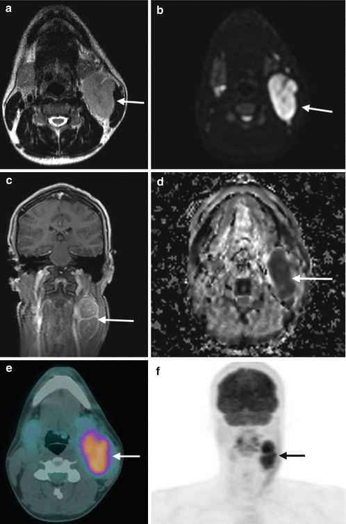
Whole-body MRI is an emerging imaging modality which holds considerable promise for staging and treatment response assessment in lymphoma [9, 10] with whole-body diffusion-weighted imaging offering functional information (by inferring cellularity) and may offer a viable alternative to (PET) CT (Fig. 4.2). Current studies of role in lymphoma imaging are mainly limited to small or pilot series, with much larger studies needed for validation.


Fig. 4.2
Whole-body MRI study in a 26-year-old male patient presenting with cervical lymphadenopathy confirmed to represent Hodgkin lymphoma by biopsy; (a) axial T2 demonstrating left deep cervical level 2/3 confluent nodal mass; (b) diffusion-weighted axial (b1000) shows high signal; (c) mDixon T1 (in phase) coronal post intravenous contrast shows moderate lesional enhancement; (d) ADC map demonstrates low signal which together with DWI imaging defines restricted diffusion suggesting hypercellularity. Correlative contemporaneous FDG PET/CT, (e) fused PET/CT images, and (f) anterior coronal maximum intensity projection (MIP) show the same lesion that demonstrates intense FDG uptake (SUVmax 10)
Integrated PET/MRI systems are becoming available for integration into routine clinical practice and offer true multifunctional imaging complemented by the molecular information of PET.
Key Points
Computed tomography (CT) has traditionally been the mainstay staging modality though increasing evidence indicates superiority of combined PET/CT in initial staging.
Stay updated, free articles. Join our Telegram channel

Full access? Get Clinical Tree


