19 Benign Enlargement of Subarachnoid Spaces
19.1 Case Presentation
19.1.1 History
A 14-month-old female patient presents with a history of macrocephaly and emesis.
19.2 Imaging Analysis
19.2.1 Imaging Findings and Impression
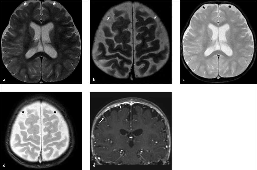
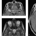
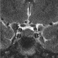
Brain MRI axial T2-weighted (T2w; ▶ Fig. 19.1a,b), axial T2*GRE (gradient recalled echo; ▶ Fig. 19.1c,d), and coronal T1-weighted (T1w) with gadolinium (▶ Fig. 19.1e) images show prominence of the subarachnoid spaces overlying the anterior cerebral convexities (asterisks) consistent with benign enlargement of subarachnoid spaces (BESS) of infancy, which is a normal variant. Note the presence of small bridging veins crossing the subarachnoid spaces (white arrow in ▶ Fig. 19.1e). Lack of susceptibility signal from blood degradation products on T2*GRE images (▶ Fig. 19.1c,d) further supports diagnosis of BESS.
19.3 Differential Diagnosis
BESS:
Normal transient enlargement of the subarachnoid spaces associated with macrocephaly.
Chronic subdural hematoma:
It should be suspected if the collection thickness is wider than 6 mm, shows higher T2 fluid-attenuated inversion recovery (FLAIR) signal as compared to cerebrospinal fluid (CSF), and displays hemorrhagic staining on T2*GRE. 1
The collection is usually asymmetric, displaces the bridging veins toward the brain surface, and has mass effect.
Cerebral volume loss/brain atrophy:
When present, it is usually accompanied by small head circumference, and “pointed” forehead due to early metopic suture fusion.
Benign enlargement of subarachnoid spaces causes an increase of the head circumference and a flat forehead due to frontal bossing.
Acquired communicating hydrocephalus:
It usually is caused by a hemorrhagic, inflammatory, or neoplastic process.
Some congenital causes include achondroplasia due to narrow foramen magnum and jugular foramina.
Density or signal intensity of extra-axial collection does not follow the CSF.
19.4 Diagnostic Pearls
Widening of the vertical distance between calvarium and brain frontal parenchyma ≥ 5 mm.
It is usually accompanied by enlarged cisterns (suprasellar and suprachiasmatic) and mildly enlarged ventricles (66%).
Normal veins are traversing subarachnoid space, the fluid in subarachnoid space follow the CSF signal on all sequences, and there is no abnormal meningeal enhancement. 2 , 3 , 4 , 5 , 6
Related posts:

Stay updated, free articles. Join our Telegram channel

Full access? Get Clinical Tree


