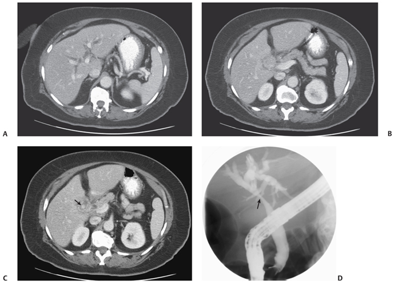
CASE 19 A middle-aged woman presents with right upper quadrant abdominal pain and jaundice. Fig. 19.1 (A–C) Axial contrast-enhanced CT images show dilatation of the intrahepatic bile ducts. There is a hyperdense stone (arrow) seen within the cystic duct associated with wall thickening. (D) Endoscopic retrograde cholangiopancreatography image shows a smooth extrinsic mass effect on the common hepatic duct (arrow). Axial contrast-enhanced computed tomography (CT) image shows dilatation of the intrahepatic bile ducts. There is a hyperdense stone seen within the cystic duct associated with wall thickening. Endoscopic retrograde cholangiopancreatography (ERCP) image shows a smooth extrinsic mass effect on the common hepatic duct (Fig. 19.1). Mirizzi syndrome (partial obstruction of the common hepatic duct due to a gallstone impacted in the cystic duct)
Clinical Presentation
Radiologic Findings
Diagnosis
Differential Diagnosis
Related posts:


Stay updated, free articles. Join our Telegram channel

Full access? Get Clinical Tree


