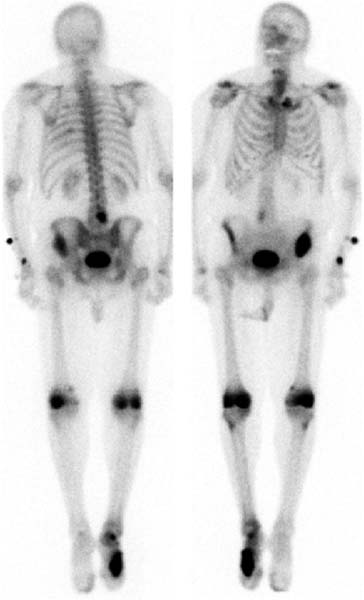
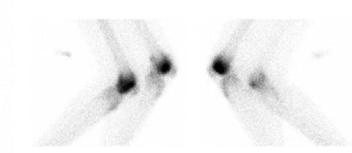
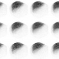
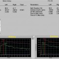
CASE 19 A 78-year-old man with a history of colon cancer presents with knee pain (Figs. 19.1 and 19.2). Fig. 19.1 Fig. 19.2 • A 20 mCi dose of 99mTc-MDP is administered intravenously. • Whole-body or spot images of the skeleton are obtained 3 hours after tracer administration. • Lateral spot views of the knees are obtained. • Emphasize the importance of oral hydration to improve soft tissue and bladder clearance. Whole-body images (Fig. 19.1) and spot views of the knees (Fig. 19.2) show mildly increased activity posteriorly in approximately the eighth left rib; in addition, intense, focally increased tracer uptake is seen in the lumbar spine at approximately L4 and L5, in the region of the anterior iliac crest on the left, in the distal femora bilaterally, and in the midfoot on the right. The injection site is noted at the intravenous access in the left distal forearm. • Metastatic disease • Trauma • Degenerative joint disease • Paget disease • Fibrous dysplasia • Avascular necrosis (of the knees) • Skin contamination (particularly at the foot)
Clinical Presentation
Technique
Image Interpretation
Differential Diagnosis
Diagnosis and Clinical Follow-Up
Stay updated, free articles. Join our Telegram channel

Full access? Get Clinical Tree


