19 The Coronary Veins
Introduction
The literature for coronary veins, in contrast to that for coronary arteries, is scarce. Moreover, a complete, highly efficient and clinically useful classification of the coronary venous system (CVS) is not as straightforward as in the case of the coronary arteries. Recent developments in cardiac pacing and transvenous ablations ask for a thorough and a more detailed knowledge of the cardiac venous anatomy and function, concomitantly demonstrating the increasing value of CVS imaging. This holds true especially in the case of cardiac computed tomography (CT) venous mapping and justifies the content of this chapter. Imaging information about the CVS in this chapter is useful for better understanding of the spatial orientation of the CVS and helps in the proper use of the correct nomenclature for this important system. The variations, anomalies, and modifications encountered in congenital heart disease are subsequently presented. We also describe clinical applications of this system in cardiac resynchronization therapy (CRT) and other interventions such as percutaneous mitral annuloplasty (PMA) and retrograde cardioplegia perfusion (RCP).
Development of the Cardiac Veins
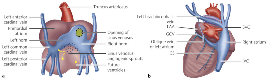
The right and left common cardinal veins (ducts of Cuvier) are formed by the confluence of the anterior and posterior cardinal veins. 1 , 2 The right horn of the sinus venosus and the right common cardinal vein will develop and eventually form the posterior wall of the right atrium (RA) and the superior vena cava (SVC), respectively. The left horn of the sinus venosus, in conjunction with the regressing left common cardinal vein, forms the coronary sinus (CS) and the ligament or vein of the left atrium (LA; Marshall; Fig. 19‑1). 1 , 2 A vessel connecting the right and left superior cardinal veins enlarges and becomes the left brachiocephalic vein. The right sinus valve persists as the valve of the inferior vena cava (IVC; Eustachian valve) and the valve of the CS (Thebesian valve). 2
It was suggested in earlier experiments that coronary vessels arise from the proepicardium, a transitory structure in the embryonic heart that forms epicardium and several internal tissues. 3 Recent histological analysis in mouse, and cardiac organ culture has shown that coronary vessels arise from angiogenic sprouts of the sinus venosus, the major vein that returns circulating blood to the embryonic heart. 4 Some sprouting venous endothelial cells dedifferentiate into arteries and capillaries as they invade the myocardium and some remain on the surface and differentiate into veins. 4
Classification of the Coronary Veins
The coronary veins depict a very different macroscopic disposition, as compared to the coronary arterial system, and show many more individual variations. Modern anatomical classification divides the cardiac veins into two major groups. These two groups include tributaries of the greater CVS and the smaller CVS, consisting of the Thebesian vessels. 5 , 6 , 7 For clarity, the greater CVS is divided into two groups: the CS and non-CS tributaries (Fig. 19‑2). Branches of the two systems can communicate. The term compound CVS has been used to describe structures in which venous drainage is performed equally by both systems (Fig. 19‑2). 6 In the ventricular myocardium, the external two-thirds are drained by the greater CVS and the internal third is mainly drained by tributaries of the smaller CVS. 6
The left ventricle (LV) and part of the right ventricle (RV) and the LA are drained by the CS tributaries and the majority of the RV and both atria are drained by the non-CS tributaries. Almost all veins of the greater CVS are finally drained into the right atrium. In the right atrium, the wall of sinus venosus is drained by tributaries of the greater CVS and the right atrial appendage is mainly drained by the smaller CVS. 8

Smaller Cardiovascular System (Thebesian Vessels)
Consisting of sinusoids, channels, and lacunae with variable sizes, this subendocardial and intramyocardial bidirectional communicating network runs in every direction conducting blood from epicardial coronaries into the cardiac chambers and provides myocardial nourishment (Fig. 19‑3 a).
The vessels of the smaller CVS are collectively called Thebesian veins. However, in view of their arterial component, the term Thebesian vessels is preferred. 6 Four distinct parts are described (Fig. 19‑3 b). Their orifices are found in cardiac chambers (mostly RA and RV) and the bases of the papillary muscles 9 , 10 and usually are greater than 0.5 mm in diameter but can be much larger especially in congenital heart disease with abnormal drainage of the CS. 11

In adults, venous drainage of the myocardial wall takes place during systole. Approximately 70 to 80% of the left coronary artery blood flow drains through the CS tributaries and the remainder is drained via the smaller CVS. The right heart chambers mainly drain thorough the smaller CVS. 6 , 8
This network can provide reverse flow from ventricles into the epicardial coronary arteries when their flow is compromised. However, it may be difficult to fill sinusoids directly from the heart chambers due to the existence of the monocuspid valve or sphincter at the outlet of the Thebesian veins. 12 It is shown that the Thebesian veins are increased in area with chronic myocardial infarction. In patients with congestive heart failure, congestion predominantly involves the large veins not the Thebesian veins. 13
Thebesian Sinusoids of Special Structures
Sinusoids are large capillaries with different morphologies. They are remnants of the primitive cardiac circulation 9 (Fig. 19‑4).

Conduction System
Both atrioventricular and sinoatrial nodal zones are sinusoid rich for retrograde blood flow to nodal conducting tissue 14 in case the arterial supply is compromised.
Papillary Muscles
The papillary muscles are mainly drained by the SVC in the RV and by the great cardiac vein (GCV) and SVC in the left. 6 Retrograde vascularization by sinusoids can supplement normal flow to papillary muscles and may prevent their rupture after ischemic damage. 15 Thebesian vessel foramina are common at the apex of the ventricles and at the base of the papillary muscles (Fig. 19‑5).

Left Ventricle Venoluminal Thebesian Vessels
A greater number of Thebesian veins are seen in the RV than in the LV. 10 Arterioluminal vessels are identified only in the RV, whereas venoluminal vessels are present in both ventricles.
Atrial Venous System
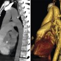
Imaging of these veins is difficult because of the small size of the vessels. However, careful evaluation of cardiac CT images can show these structures in many studies. Different classifications have been introduced. 6 , 8 , 16 , 17 The easiest classification is introduced by von Lüdinghausen 8 (Table 19‑1).
Veins of the Left Atrial Wall
The majority of the left atrial (LA) veins are tributaries of the greater CVS. Three groups are described 6 , 8 :
Veins of the posterior and lateral walls of the LA empty into the CS and GCV (Fig. 19‑6).
Septal veins of the LA drain into the RA through the interatrial septum. These veins are commonly seen with CT angiography (CTA). Veins anterosuperior to the fossa ovalis pass through the interatrial groove and empty into the right atrium near the superior cavoatrial junction (Fig. 19‑7). Some (12%) may pass through the superior interatrial muscle connections where Bachmann’s bundle is located 18 (Fig. 19‑8). Veins inferoposterior to fossa ovalis are less common (Fig. 19‑7).
The posterosuperior wall of the LA near the LA appendage drains into the LA and the superior pulmonary veins (Fig. 19‑7). They may connect mediastinal veins, azygos, or hemiazygos venous system, and the bronchial veins. 8 The mediastinal connections can be an important alternative route for venous return to the atria in case of occlusion of the pulmonary vein, brachiocephalic vein, or SVC (Fig. 19‑7 , Fig. 19‑9). In congenital atresia of the CS, the myocardial venous drainage can be via ectatic atrial veins of the LA. 19

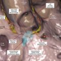
Fig. 19.6 (a) Atrial venous system. Posterosuperior view of the heart is depicted. The right atrium is removed. (1) Veins of the posterior and lateral walls of the left atrium (LA) empty into the coronary sinus (CS); (2) septal veins of the LA drain into the right atrium (RA); (3) posterosuperior wall of the LA drains into the LA lumen or the superior pulmonary veins; (4) atrioventricular node region contains small sinusoids draining into the RA near the CS ostium; (5) anterior cardiac veins may drain directly into the RA, small cardiac vein, or into a venous tunnel in the RA wall. (b) Right lateral view showing drainage of the anterior cardiac vein (ACV) into the venous tunnel running (green arrows) parallel to the atrioventricular groove. GCV, great cardiac vein; IVC, inferior vena cava; LMV, left marginal vein; LV, left ventricle; OVM, oblique vein of Marshall; RV, right ventricle; SVC, superior vena cava. 
Fig. 19.7 (a) Septal views of the right atria (RA) and the left atria (LA) from two different cadaveric specimens are shown. Most of the septal venous drainage occurs from the LA into the RA. Venous drainage of the anterosuperior and posteroinferior of the interatrial septum and posterosuperior wall of LA and atrioventricular node (AVN) region are shown by green, blue, white, and pink circles, respectively. Most of the septal venous drainage occurs from the LA into the RA. The venous opening can be large enough to be detected by CT scan. (b) Short-axis CT images demonstrate the drainage pattern of atrial wall veins matching to the colored circles shown in (a), including communication between the two atrial walls near the SVC (green arrow in b1) and IVC (blue arrow in b3), the superior wall of the LA near confluence with the left superior pulmonary vein (white arrow in b2), and the medial RA wall near the coronary sinus (CS) ostium (pink arrow in b4). Veins of the superior wall of LA may communicate with venous collaterals in the mediastinum (white arrow in b2). FO, fossa ovalis; IVC, inferior vena cava; LAA, left atrial appendage; SVC, superior vena cava; TV, tricuspid valve. 
Fig. 19.8 Venous drainage of the anterosuperior septal wall of left atrium (LA) into the right atrium (RA) through the superior interatrial muscle connection where the Bachmann bundle resides. Communications found in two different patients are shown by pink circles (a, b) and green circles (c, d). This communication is seen in 12% of CT angiographies. The lumen of a patent foramen ovalis (PFO) is incidentally seen (white arrows). SVC, superior vena cava. 
Fig. 19.9 (a) Coronal and (b) sagittal views demonstrate drainage of the collateral mediastinal veins (arrows) into the superior wall of the left atrium (LA) in a patient with chronic thrombosis of the left brachiocephalic vein. Note ascending aortic (AA) stent graft in the area of aneurysm. SVC, superior vena cava.
Veins of the Right Atrial Wall
Majority of the right atrial wall veins are located intramurally and drain into the RA. The veins draining the wall of the right atrial appendage have no connection with the epicardial veins, and therefore are classified as smaller CVS. The RA veins are classified into four groups based on anatomical locations (Table 19‑1):
Small intramural venous tunnels draining directly into the RA. They are a few millimeters in length and 1 mm in diameter. 6
Venous tunnels of the RA (the sinus coronaries atrii dextri or right atrial CS): Termination of the anterior cardiac veins of the RV is variable. They may drain directly into the RA, small cardiac vein, or in some into a venous tunnel. 6 When it exists, this venous tunnel runs parallel and 1 to 2 cm above the tricuspid orifice along the posterior or posterolateral wall of the RA 20 (Fig. 19‑6). In high-quality CTAs, these veins may be partially visible (Fig. 19‑6 b).
The sinoatrial node region has sinusoids draining into the RA. 14 This network of anastomosing blood vessels potentially can protect the node in case arterial supply is compromised. However, this phenomenon is rare given the common variants of dual or multiple arterial supplies to the node.
Atrioventricular node region: Venous tunnels are seen at the base of the interatrial septum near the atrioventricular node and the bundle of His and usually open into the RA near the ostium of the CS or directly into the CS (Fig. 19‑6 , Fig. 19‑7). These tunnels can be seen in CTA and should not be confused with anomalous vessels or a patent foramen ovale.
Anterior Right Ventricle Venous System
Including the anterior cardiac, right marginal, and conus veins are seen in 90% of cases 6 , 20 , 21 (Table 19‑2). The anterior cardiac veins drain two-thirds of the RV including the anterior and anterolateral walls of the RV. These veins are frequently seen in coronary CTAs (Fig. 19‑10). They are variable in sizes and numbers and usually drain into the RA above the atrioventricular groove. 6 , 16 , 21 , 22 In 5 to 27%, they merge to form a common trunk before entering the RA. 6 , 21 The right marginal vein originates near the right ventricular apex and receives branches from posterior and anterior surfaces of the RV (Fig. 19‑10). In one-third of cases, the vein continues as small cardiac vein into the CS. 6 In the remainder of cases, it drains into the RA or the venous tunnels of the RA.
The conus (infundibular) veins are usually small veins and drain into the RA. However, they can be large and varicoid (i.e., arteriovenous fistula) creating diagnostic problems (Fig. 19‑11) or causing excessive bleeding during surgery. Veins of Zuckerkandl and Cruveilhier are described in the anatomic literature for veins draining the anterior and posterior surfaces of the infundibulum, respectively 20 , 21 (Fig. 19‑11).


Coronary Sinus Tributaries
The CS tributaries include the GCV, the left marginal vein, the posterolateral vein of the LV, and the inferior interventricular vein (IIV; Fig. 19‑12). In 30% of cases, it receives the right marginal vein blood through the small cardiac vein 6 (Table 19‑2).

Imaging Methods
The CS is frequently used as a conduit for catheter treatment of arrhythmias as well as left ventricular pacing. These procedures are well known for their potential technical difficulties. As a result, different imaging methods are introduced for evaluation of this system. Each technique has its own advantages and limitations. 23 , 24 , 25 , 26 , 27 , 28 , 29
Retrograde venography via the CS is currently the standard technique for defining the CS anatomy; however, it is invasive in nature and can be technically challenging. This technique is frequently used to show the anatomy of the CS and target veins for CRT. In this technique, using an occlusive balloon, contrast is injected into the CS and two orthogonal views including right anterior oblique 45 degrees and left anterior oblique 45 degrees are obtained. 23 High-speed rotational coronary venography uses rapid isocentric rotation over a 110-degree arc in 4 seconds, acquiring 120 frames per angiogram. 23 With this modification, vessel overlap and foreshortening will be reduced and target veins including second-degree tributaries can be better defined.
Levophase of coronary angiography is preferred by some investigators and claimed to better define the anterior (superior) interventricular vein (AIV) and IIV as well as the peripheral branches compared to retrograde venography. 24 The technique takes advantage of the coronary artery angiography procedure commonly indicated in the assessment of candidates for CRT.
Magnetic resonance imaging (MRI) and CT have been increasingly used to assess anatomy of the coronary vessels including coronary veins. 25 , 26 , 27 , 28 , 29 CT has the advantage of showing part of the CS and in some cases the origin of the IIV, which will be obscured with balloon in retrograde venography. With CT, there is no foreshortening or vessel overlap. CT also demonstrates many ancillary structures such as coronary arteries, old infarctions, interatrial septum anatomy, and variants of the normal anatomy. Higher radiation dose in CT is a limitation 26 that can be modified using dose reduction techniques. CT also may have difficulty showing second-order tributaries of the coronary veins in routine coronary CTAs. Demonstration of the oblique vein of LA is also more challenging with CT compared with retrograde venography. However, with some modification of the CT technique, most small branches can be nicely depicted. 18
Great Cardiac Vein
The GCV is the longest and the most consistent vessels making up the cardiac venous system, almost always entering the CS. 30 , 31 , 32 , 33 , 34 This vein drains blood from the anterior interventricular septum, the anterior surfaces of both ventricles, part of the LA, and the apical region of the heart. 6 The AIV portion of the vein courses superiorly within the anterior (superior) interventricular groove (sulcus) and then enters the left atrioventricular groove where it is defined as the GCV. The GCV may originate at the apex or in the apical or middle third of the anterior interventricular groove with a highly variable position with respect to the left anterior descending (LAD) artery, which may render very difficult the localization of the latter especially in coronary artery bypass procedures.
The GCV crosses the branches of the left coronary artery system including the anterior descending and the circumflex branches forming a triangle 6 , 30 in which the vein is mainly superficial to arteries in 60 to 70% 31 , 32 , 33 , 34 of cases (Fig. 19‑13). Branches of the left coronary artery system traverse this triangle (i.e., the ramus intermedius). However, it is important to know that the relation of the vein and these arteries is highly variable and practically unpredictable in many cases.
Aberrant drainage of the GCV into the RA or SVC is described. 33 In this situation, the GCV runs in the transverse sinus and finally joins the azygos vein or the RA. Knowledge of this anatomy prior to surgery can be important for RCP.
Myocardial bridges are detected above the AIV or its tributaries in 8%. 30 Aneurysmal dilatation of the distal end of the GCV has been reported and may interfere with coronary artery bypass grafting (CABG). 34 It is the second most common location of coronary vein aneurysm seen in 1.5% of CT examinations 35 (Fig. 19‑14).


Inferior Interventricular Vein
The IIV, also known as middle cardiac vein or posterior interventricular, travels along the inferior aspect of the heart (attitudinal orientation) in the interventricular groove. The vein is a constant landmark and drains into the CS approximately 1 cm from the CS ostium. Extending from the apex to the CS, it drains the interior walls of the ventricles as well as the apical area and the posterior two-thirds of the septum 6 (Fig. 19‑15). The terminal portion of the vein measures 3 to 4 mm in diameter and appears angled in 35% or bulbous in 19% of population. 6 Fusiform aneurysmal dilatation (focal dilatation of twice the normal diameter; mean; 10.5 ± 1.4 mm) of the venous confluence is reported in CT studies in 8% (Fig. 19‑15). It is the most common location for bulbous enlargement of the coronary veins. 35 Aneurysms, especially the diverticular form, arising from the CS and the confluence of coronary veins with the CS, may be associated with cardiac arrhythmias such as ventricular preexcitation. 36 , 37

Left Posterior (Posterolateral) and Left Marginal Veins
Several venous branches drain the myocardial walls located between the IIV and the AIV. 30 Veins draining the posterior and lateral walls of the LV are of particular interest for the LV pacing in CRT 38 (Fig. 19‑16). Imaging techniques permit identification of potential anatomic factors that may pose difficulties to cannulate and advance the LV pacing lead including ostial valves, variceal veins, or absence of the posterior or lateral vein.

The left posterior (inferior) veins are one to three in numbers and measure 2.5 mm at ostium. While the distal myocardial origin of the left posterior vein is highly variable, it usually terminates in a uniform fashion in the CS (75%), although it can also drain into the GCV. 6 , 32 , 39 It is single in 60%. In the absence of the left posterior vein (5%), it is the left marginal vein that drains most of the left ventricular posterolateral wall. 5 Small diameter of the vein (<2 mm) and acute angulation with the CS may complicate the insertion of the lead for CRT. 38
The left marginal (obtuse marginal) vein is present in 70 to 95% of specimens, emptying into the GCV in 80% of cases and into the CS in the remaining. 30 The frequency of visualization is less with CT or MRI compared with retrograde venography. 25 , 26 , 27 , 28 It drains much of the LV myocardium, usually running superficial to the marginal branch of the circumflex artery. 32 , 40 Its diameter varies and, like other coronary veins, is enlarged in patients with cardiac failure. 40 Anastomosis of the epicardial veins in the periphery is common. This is especially common between the veins of the LV such as between the GCV and posterolateral vein or between the IIV and posterolateral or marginal veins (Fig. 19‑12).
Small Cardiac Vein
With a frequency of 30 to 50% and average diameter of 1 mm, the small cardiac vein (right coronary vein) drains the posterolateral wall of the RV into the CS (85%), the IIV (12%), or rarely the RA (1%) 16 , 30 , 41 (Fig. 19‑12). It is more common in women than in men. 41
Oblique Vein of the Left Atrium (Marshall)
The oblique vein of Marshall (OVM), a remnant of the left SVC, descends along the lateral and inferior walls of the LA between the LA appendage and the left pulmonary veins and joins at the junction of the GCV and the CS approximately 3 cm from the CS ostium. 42 , 43 The vein is short (2–3 cm in length) and its superior part obliterated by fibrosis. Complete fibrosis or obliteration in the form of a cord is seen in 5 to 12%. 6 The average diameter is 1 mm (0.4–1.8) and the angle with the CS varies between 25 and 50 degrees. It is present in 85 to 95% 42 of cases (Fig. 19‑16). Balloon-occluded venography of the CS can visualize the OVM in 73% of the patients. 44 Demonstration of this vein with CT requires special attention to the detail and quality of the CTA. It is reported in 35 to 40% of cases 25 , 39 (Fig. 19‑15 c, Fig. 19‑16 a). The best phase for visualization of the OVM is ventricular systole, likely due to coronary vein enlargement during this phase. 39
The OVM is now implicated as a source of activation in atrial tachyarrhythmias. 45 , 46 , 47 , 48 , 49 This may originate from the muscle sleeve of the vein (Marshall’s bundle). 50 This muscle sleeve is connected to the musculature surrounding the CS or the left pulmonary veins 45 , 46 , 47 (also see section “Coronary Sinus Interatrial Muscle Connections”). The vein is of greater significance when it persists as a patent left SVC. It is seen in 0.5% of the normal population and up to 5% of congenitally malformed hearts 51 draining into the CS.
Related posts:

Stay updated, free articles. Join our Telegram channel

Full access? Get Clinical Tree


