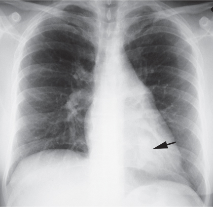
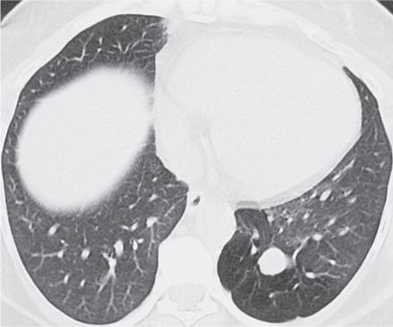
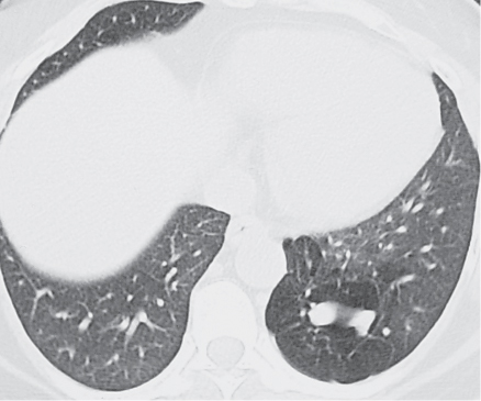
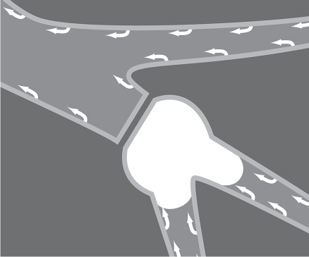
CASE 2 Asymptomatic 42-year-old woman PA (Fig. 2.1) chest radiograph demonstrates a left lower lobe branching tubular opacity (arrow). Chest CT (lung window) (Figs. 2.2, 2.3) shows the branching tubular lesion (Fig. 2.3) surrounded by hyperlucent lung. Artist’s illustration (Fig. 2.4) shows the proposed etiology of bronchial atresia: an interruption of the bronchial lumen produces a mucocele distal to the atresia formed by accumulation of secretions (carried centrally by the mucociliary escalator). Bronchial Atresia • Mucoid Impaction Distal to Bronchial Obstruction (e.g., endobronchial neoplasm) • Focal Bronchiectasis with Mucoid Impaction • Allergic Bronchopulmonary Fungal Disease • Vascular Malformation • Intralobar Sequestration Fig. 2.1 Fig. 2.2 Fig. 2.3 Fig. 2.4
 Clinical Presentation
 Radiologic Findings
 Diagnosis
 Differential Diagnosis
 Discussion
Background
Related posts:
![]()
Stay updated, free articles. Join our Telegram channel

Full access? Get Clinical Tree


Radiology Key
Fastest Radiology Insight Engine


