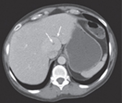
CASE 2 A 42-year-old woman presents with upper right quadrant tenderness and hepatomegaly. Fig. 2.1 Abdominal contrast-enhanced CT scan shows an ill-defined, homogeneously enhancing lesion within the caudate lobe of the liver (arrow) with a hypodense central scar (dashed arrow). Abdominal computed tomography (CT) scan (Fig. 2.1) reveals a poorly defined lesion within the caudate lobe with a low-density central scar. Abdominal magnetic resonance imaging (MRI) (Fig. 2.2) was subsequently performed. The liver mass appears isointense to normal liver parenchyma on T2- and T1-weighted images with a central scar that is T2 hyperintense. Arterial enhancement is noticed within the lesion as well as delayed enhancement of the scar. Focal nodular hyperplasia (FNH) FNH is the second most common hepatic benign tumor after hemangioma and is found in 1% of the population (mainly middle-aged women). It is most frequently seen as a solitary lesion even though multiple masses have been described in 20% of patients. Histologically, FNH is characterized by the presence of normal hepatocytes with a malformed biliary draining system. Prompt diagnosis is mandatory to distinguish this lesion from malignant hepatic masses such as fibrolamellar carcinoma, HCC, cholangiocarcinoma, and hypervascular metastasis.
Clinical Presentation

Radiologic Findings
Diagnosis
Differential Diagnosis
Discussion
Background
Stay updated, free articles. Join our Telegram channel

Full access? Get Clinical Tree


