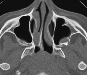
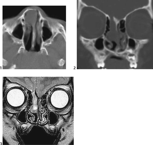
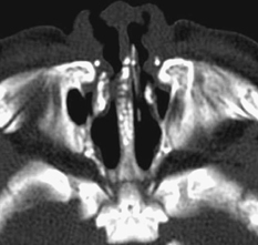
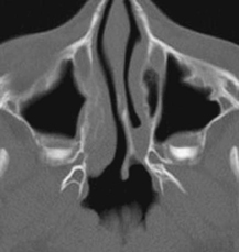
CASE 20 A 5-day-old newborn was sent for imaging due to failure to pass a nasogastric tube on the right side. Figure 20A Axial CT scanning (Fig. 20A) shows a funnel-shaped posterior portion of the right nasal cavity. An abnormal medial deviation of the right lateral nasal wall and perpendicular palatine bone with an expanded medial pterygoid plate with a widened vomer is also seen. Soft tissue plug fills the right nasal cavity just in front of the ipsilateral closed choana. Figure 20B The following figures demonstrate right frontonasal encephalocele. Axial CT (1) at nasal cavity of a right frontonasal encephalocele is shown as a soft tissue mass immediately to the right of the midline in the anterior nasal cavity. The coronal CT (2) delineates the bone defect at the anterior portion of the right cribriform plate. The coronal MRI T2-weighted image (3) shows herniation of the right gyrus rectus and olfactory bulb through the defect into the nasal cavity. Choanal atresia, unilateral The etiologic diagnosis of neonatal nasal obstruction includes: Neonates are obligate nasal breathers and nasal obstruction, especially if bilateral and complete, may cause severe respiratory distress. The posterior choanae are the orifices that connect the posterior nasal cavity with the nasopharynx. Atresia or narrowing of the posterior choanae is the most common cause of nasal obstruction in the neonate, with an incidence of 1 in 5000 to 8000 births. Figure 20C Nasal aperture stenosis was observed in a newborn. Axial CT of the nasal bone (angled along the hard palate) shows dysmorphic features, sacral dimple, and chondrodysplasia punctata type. There is narrowing of the piriform aperture and of the anterior nasal cavity. The maxillary spines show abnormal punctate calcification pattern. Figure 20D Membranous type choanal atresia in a 5-year-old child. Axial CT shows slight mucosal thickening anterior to the band of left choanal atresia.
Clinical Presentation
Radiologic Findings
Diagnosis
Differential Diagnosis
Discussion
Background
Related posts:


Stay updated, free articles. Join our Telegram channel

Full access? Get Clinical Tree


