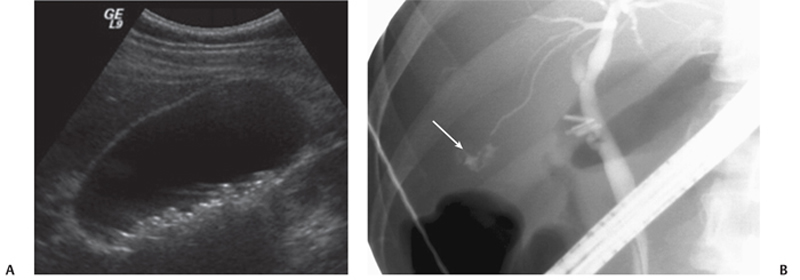
CASE 22 A 37-year-old woman presents with fever and abdominal pain 2 days following a cholecystectomy. Fig. 22.1 (A) Ultrasound image obtained prior to cholecystectomy demonstrates multiple stones layering posteriorly within the gallbladder. (B) Image from an endoscopic retrograde cholangiopancreatography obtained 2 days following laparoscopic cholecystectomy demonstrates free contrast extravasation (arrow) into the gallbladder fossa through an accessory hepatic duct fed from a left hepatic duct branch. An ultrasound image obtained prior to the cholecystectomy demonstrates multiple stones layering posteriorly within the gallbladder. An image from endoscopic retrograde cholangiopancreatography (ERCP) obtained 2 days after a laparoscopic cholecystectomy demonstrates free contrast extravasation into the gallbladder fossa through an accessory hepatic duct fed from a left hepatic duct branch (Fig. 22.1). Bile leak secondary to bile duct injury For perihepatic fluid following hepatobiliary surgery seen on imaging:
Clinical Presentation
Radiologic Findings
Diagnosis
Differential Diagnosis
Discussion
Background
Related posts:


Stay updated, free articles. Join our Telegram channel

Full access? Get Clinical Tree


