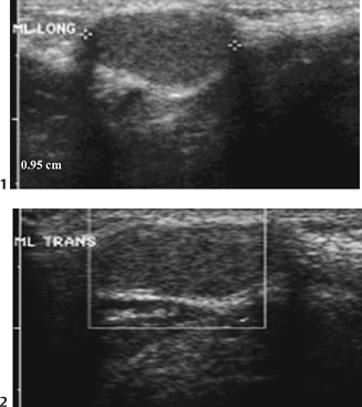
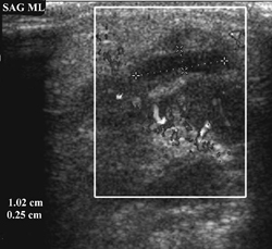
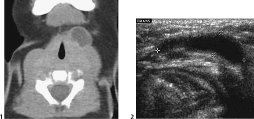
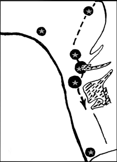
CASE 22 A young child presents with a midline enlarging neck lump just below the chin, above the level of the hyoid bone. Figure 22A Ultrasonography of the anterior neck in the sagittal (Fig. 22A1) and transverse (Fig. 22A2) planes demonstrates a well-defined midline echogenic mass just above the hyoid bone. Figure 22B Midline sagittal sonogram in submental area demonstrating early abscess formation and adjacent inflamed lymph node. Figure 22C Left anterior cystic hygroma of the neck. Axial CT (1) showing a well-defined hypodense cyst. Corresponding transverse sonogram (2) demonstrates a superficial off-midline anechoic cyst. Thyroglossal duct cyst The most common congenital neck anomaly, thyroglossal duct cysts, accounts for 70% of congenital neck anomalies and are frequent neck masses second only to benign lymphadenopathy. It has equal incidence in males and females. Seven percent of the adult population may have such a remnant. Familial cases are rare but do occur, and they may be autosomal dominant or recessive. Figure 22D Diagram showing possible locations (stars) of thyroglossal duct cysts along their embryologic path. Landmarks are the foramen cecum of the tongue base, the hyoid bone, the thyroid cartilage, and the sternum.
Clinical Presentation
Radiologic Findings
Diagnosis
Differential Diagnosis
Discussion
Background
Embryology
Related posts:


Stay updated, free articles. Join our Telegram channel

Full access? Get Clinical Tree


