III Pancreas
CASE 23
Clinical Presentation
A 20-year-old woman with a clinical history of recurrent episodes of pancreatitis presents with abdominal pain.
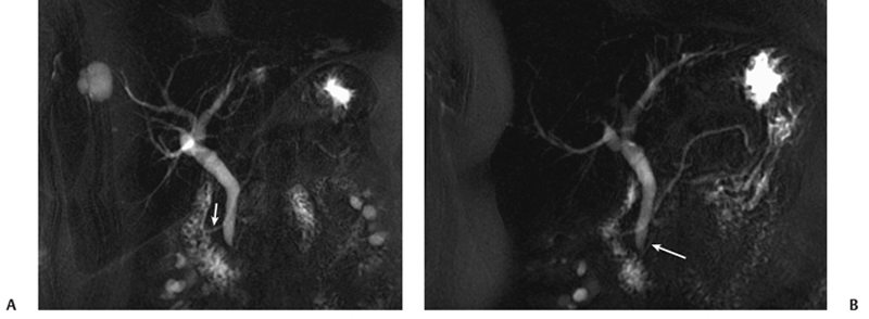
Fig. 23.1 (A) Magnetic resonance cholangiopancreatography (MRCP) image shows the longer dorsal duct of Santorini (arrow) draining into the minor papilla. (B) MRCP image at a different level shows the smaller ventral duct (arrow) joining the common bile duct and draining into the major papilla. Note the dorsal duct crossing over the common bile duct, which is a characteristic MRCP appearance in this congenital anomaly.
Radiologic Findings
Magnetic resonance cholangiopancreatography (MRCP) images (Fig. 23.1) show a longer dorsal pancreatic duct (duct of Santorini) draining in the papilla; the ventral duct (Wirsung duct) appears smaller and shorter, and it drains in the major papilla.
Diagnosis
Pancreas divisum
Differential Diagnosis
- None
Discussion
Background
Related posts:


Stay updated, free articles. Join our Telegram channel

Full access? Get Clinical Tree


