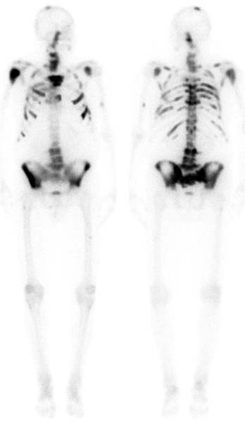
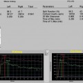
CASE 23 A 71-year-old man with history of prostate cancer undergoes bone scan to assess bony metastasis (Fig. 23.1). Fig. 23.1 • A 20 mCi dose of 99mTc-MDP is administered intravenously. • Whole-body images of the skeleton are obtained 3 hours after tracer administration. • A 1024 × 256 matrix is used for whole-body images. • Emphasize the importance of oral hydration to improve soft tissue and bladder clearance. Whole-body images of the skeleton demonstrate increased tracer uptake in many foci throughout the axial skeleton and in the vertex of the skull. There are also areas of decreased tracer activity from the midthoracic region down to approximately L3 and in the lower half of the pelvis. (Region of decreased tracer uptake) • Radiation therapy • Prosthetic implant
Clinical Presentation
Technique
Image Interpretation
Differential Diagnosis
Stay updated, free articles. Join our Telegram channel

Full access? Get Clinical Tree


