CASE 24 A 6-month-old baby girl presents with growing right facial mass. Figure 24A Figure 24B (See Color Plate 24B.) Figure 24C Figure 24D Figure 24E Ultrasonographic images show a large, soft tissue mass of heterogeneous echogenicity (Figs. 24A and 24B). Color Doppler evaluation shows high flow and high vessel density (Fig. 24B). MRI including axial unenhanced T1-weighted image (Fig. 24C), coronal fat-suppressed T2-weighted image (Fig. 24D), and fat-suppressed T1-weighted image following gadolinium enhancement (Fig. 24E) show a large mass initially isointense to muscle within the right parotid gland extending to the right parapharyngeal region with multiple internal flow voids. The lesion shows high T2 signal intensity and intense enhancement following gadolinium administration. Hemangioma of infancy Other vascular anomalies and tumors
Clinical Presentation
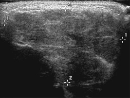
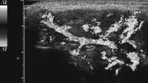
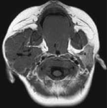
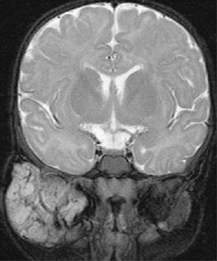
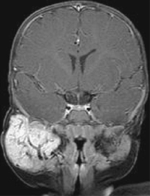
Radiologic Findings
Diagnosis
Differential Diagnosis
Discussion
Background
Related posts:


Stay updated, free articles. Join our Telegram channel

Full access? Get Clinical Tree


