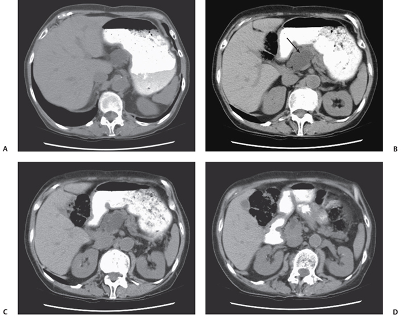
CASE 24 A 70-year-old man presents with abdominal pain and weight loss in the last few months. Fig. 24.1 (A–D) Axial unenhanced abdominal CT scans show a hypodense, well-defined, partially lobulated lesion localized in the neck of the pancreas (arrow) that appears to be in association with the main pancreatic duct, which is focally dilated and anteriorly displaced. The ampullary tract of the main pancreatic duct is not dilated, and there is no intra- or extrahepatic biliary dilatation. The body and tail of the pancreas are otherwise unremarkable and present mild fatty replacement. No lymphoadenopathy is noticed. An unenhanced axial computed tomography (CT) scan of the abdomen reveals a hypodense, multilocular, lobulated lesion localized in the neck of the pancreas (Fig. 24.1). The main pancreatic duct is mildly dilated and seems in continuity to the hypodense mass. Intraductal papillary mucinous neoplasia (IPMN) IPMN and other pancreatic cystic neoplasms have been increasingly identified over the last decade at cross-sectional imaging due to the recent introduction of dedicated thin-section CT protocol and magnetic resonance cholangiopancreatography (MRCP); thus, a diagnosis of these different entities is mandatory according to respective differences in clinical management. IPMNs have typical features, as they grow in a papillary fashion within either the main pancreatic duct or the side branches or both and produce thick mucinous secretions, causing diffuse or focal dilatation of the pancreatic ducts. They are frequently observed in elderly men and represent ˜3 to 5% of all pancreatic neoplasms and 20% of cystic pancreatic lesions.
Clinical Presentation

Radiologic Findings
Diagnosis
Differential Diagnosis
Discussion
Background
Clinical Findings
Stay updated, free articles. Join our Telegram channel

Full access? Get Clinical Tree


