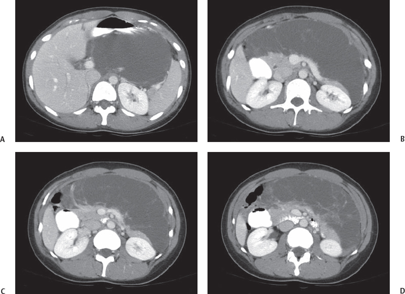
CASE 25 A 38-year-old woman presents with abdominal pain and epigastric tenderness. Fig. 25.1 (A–D) Images from a contrast-enhanced abdominal CT scan show a large, lobulated, well-defined, hypodense, finely septated mass associated with the pancreatic head and body. Abdominal contrast-enhanced computed tomography (CT) scans (Fig. 25.1) show a large, lobulated, hypodense lesion with multiple thin septations, centered in the upper abdomen, intimately associated with the pancreas. The superior mesenteric artery and vein and the portal vein are patent. Pancreatic lymphangioma
Clinical Presentation
Radiologic Findings
Diagnosis
Differential Diagnosis
Discussion
Background
Related posts:


Stay updated, free articles. Join our Telegram channel

Full access? Get Clinical Tree


