CASE 26 A 68-year-old man presents with vomiting and epigastric pain. Fig. 26.1 (A–C) Images from a contrast-enhanced abdominal CT scan reveal massive fluid collections extending in the abdomen along the anterior aspect of the liver, in the mesentery, and in the anterior pararenal fasciae. The pancreatic parenchyma shows no enhancement and is almost completely replaced by ill-defined fluid collections. Diffuse mesenteric and peripancreatic fat stranding consistent with inflammatory changes is also noted. Abdominal contrast-enhanced computed tomography (CT) scan (Fig. 26.1) shows diffuse inflammatory changes in the peripancreatic region, seen as peripancreatic and mesenteric fat stranding, extensive peripancreatic and perihepatic fluid collection, and absence of enhancement within the pancreatic head, neck, and body. Acute necrotizing pancreatitis Acute pancreatitis coexisting with one of the following:
Clinical Presentation
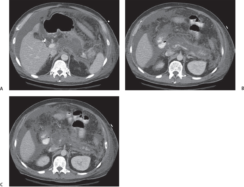
Radiologic Findings
Diagnosis
Differential Diagnosis
Discussion
Background
Related posts:


Stay updated, free articles. Join our Telegram channel

Full access? Get Clinical Tree


