CASE 26 A 55-year-old man with a history of benign prostatic hyperplasia presents with elevated prostate-specific antigen (Fig. 26.1). Fig. 26.1 • A 20 mCi dose of 99mTc-MDP is administered intravenously. • Whole-body images of the skeleton are obtained 3 hours after tracer administration. • A 1024 × 256 matrix is used for whole-body images. • Emphasize the importance of oral hydration to improve soft tissue and bladder clearance.
Clinical Presentation
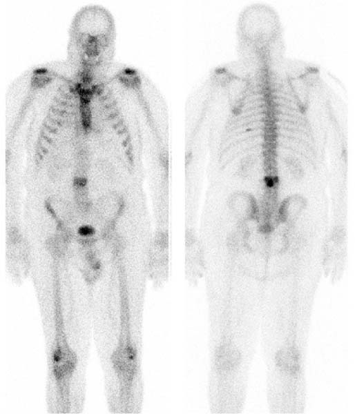
Technique
Image Interpretation
Related posts:


Stay updated, free articles. Join our Telegram channel

Full access? Get Clinical Tree


