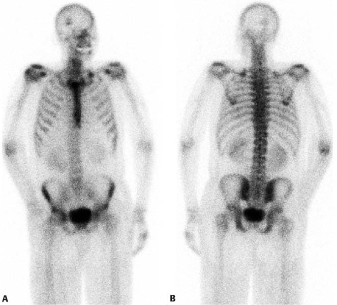
CASE 27 A 41-year-old man presents with an abnormality in his head noted on gallium scan done 1 month prior (Fig. 27.1). Fig. 27.1 • A 20 mCi dose of 99mTc-MDP is administered intravenously. • Whole-body images of the skeleton are obtained 3 hours after tracer administration. • A 1024 × 256 matrix is used for whole-body images. • Emphasize the importance of oral hydration to improve soft tissue and bladder clearance. Tracer uptake is noted in the right side of the skull in the right anterior oblique view (Fig. 27.1A). The posterior view of the skull (Fig. 27.1B) suggests a faint lesion that may be related to the occipital abnormality noted on the gallium scan or that may be shine-through of the lesion seen on the right in the anterior view.
Clinical Presentation
Technique
Image Interpretation
Related posts:


Stay updated, free articles. Join our Telegram channel

Full access? Get Clinical Tree


