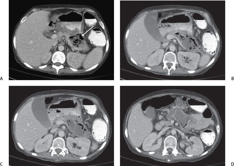
CASE 27 A 65-year-old man presents with epigastric pain. Fig. 27.1 (A–D) Contrast-enhanced axial images from an abdominal CT scan show the pancreatic parenchyma replaced by a low-density fluid collection. The collection is well defined and has mildly enhancing walls. Gas is seen within the collection (arrow). The portal and splenic veins are patent. Abdominal contrast-enhanced computed tomography (CT) scan (Fig. 27.1) shows a hypodense fluid collection replacing most of the pancreatic parenchyma and exerting mass effect on the posterior margin of the stomach. Gas is seen within this fluid collection. The portal and splenic veins are patent. Pancreatic abscess
Clinical Presentation
Radiologic Findings
Diagnosis
Differential Diagnosis
Discussion
Background
Related posts:


Stay updated, free articles. Join our Telegram channel

Full access? Get Clinical Tree


