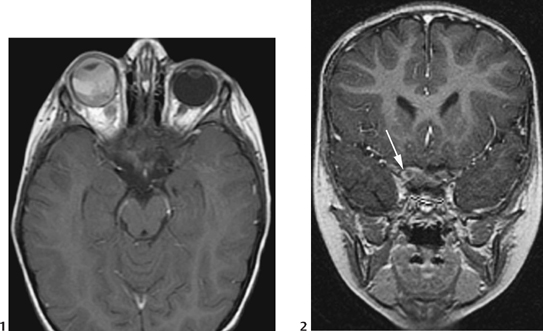
CASE 27 A 9-month-old male presents with a 4-week history of right esotropia (inward turning of the eye) and periorbital swelling. Figure 27A Figure 27B CT (Fig. 27A) reveals bilateral calcified mildly enhancing intraocular masses with right periorbital swelling. Axial fast spin echo T2-weighted MRI (Fig. 27B) demonstrates bilateral intraocular hypointense masses consistent with partly calcified retinoblastoma. Bilateral retinoblastoma Retinoblastoma is the most common intraocular tumor of childhood. It occurs in 1 in 10,000 to 15,000 live births. It is nonhereditary in 60 to 80% of cases and hereditary in the remainder. The hereditary form usually presents earlier (<2 years of age) and is frequently bilateral. Trilateral retinoblastoma represents the association of a midline intracranial tumor (usually pineoblastoma) with bilateral retinoblastoma. Retinoblastoma can also be rarely tetralateral when it is associated with a suprasellar tumor. The etiology of retinoblastoma includes both hereditary and nonhereditary forms. The gene responsible for retinoblastoma is localized to the 13q14 locus, which is mutated or deleted in both hereditary and nonhereditary forms. The retinoblastoma gene functions as a suppressor gene. If both alleles are suppressed or deleted, then retinoblastoma can develop. The hereditary form is autosomal dominant with a penetrance of 90% and with increased risk in trisomy 21. Retinoblastoma arises from retinal neuroectodermal cells. Leucocoria (white papillary reflex) or “cat’s eye” is the most common presentation, often shown on photographs as loss of the red eye. Other presentations include: Macroscopically, retinoblastomas are noted as single or multiple white, “chalky” nodules in the retina. There are three types: Figure 27C Axial (1) and coronal (2
Clinical Presentation
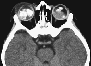
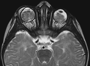
Radiologic Findings
Diagnosis
Differential Diagnosis
Discussion
Background
Etiology
Embryology
Clinical Findings
Pathology
![]()
Stay updated, free articles. Join our Telegram channel


Full access? Get Clinical Tree


