Case 28
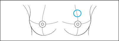
Indication: 2 cm mass between upper quadrants of the left breast.
History: Unremarkable.
Risk profile: No increased risk.
Age: 35 years.
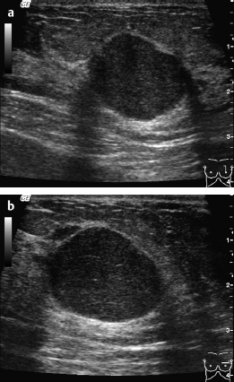
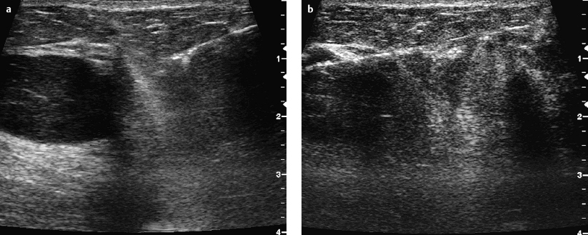
Fig. 28.1 a,b Ultrasound.
Clinical Findings
Resistance (diameter 2 cm) between upper quadrants of the left breast.
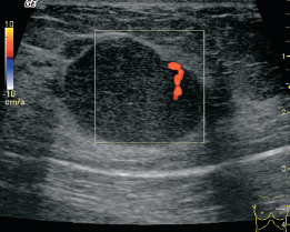
Fig. 28.2 Color-coded Doppler sonography.
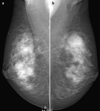
Fig. 28.3a,b Digital mammography, MLO view.
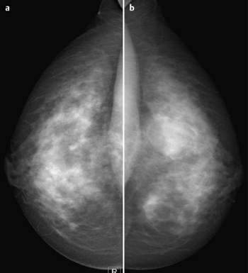
Fig. 28.4a,b Digital mammography, CC view.
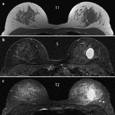
Fig. 28.5a–c Contrast-enhanced MRI of the breasts.
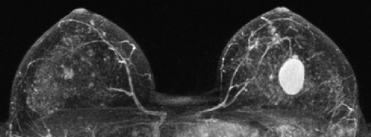
Fig. 28.6 Contrast-enhanced MR mammography. Maximum intensity projection.
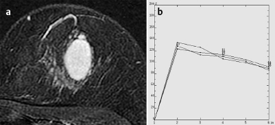
Fig. 28.7a,b Signal-to-time curves.

|
Please characterize ultrasound, mammography, and MRI findings.
What is your preliminary diagnosis?
What are your next steps? |
This case examines the imaging studies of a young woman with a recently detected resistance between the upper quadrants of the left breast.
Ultrasound
Corresponding to the resistance between the upper quadrants of the left breast, along a vertical axis from the nipple, the ultrasound depicted an oval hypoechoic lesion (diameter: 2,5 cm) with moderate distal acoustic increase and a lateral echo decrease. US BI-RADS3.
Mammography
Mammograms showed inhomogeneous dense parenchyma ACR type 3. Between the upper quadrants of the left breast an isodense, partially ill-defined lesion (diameter 2,5 cm) with a semicircular “Halo sign” was depicted. There were no other suspect densities or architectural distortions and no microcalcifications visible. BI-RADS right 1/left 3. PGMI: P for CC; M for MLO (inframammary fold).
MR Mammography
In the central region of the left breast MRI demonstrated a well-defined mass (diameter 3 cm) enclosed by a narrow section of lipomatous tissue. After administration of contrast, homogeneous enhancement with strong initial signal increase (120%) followed by washout were visible. Signal in T2-weighted imaging was increased.
MRI Artifact Category: 1
MRI Density Type: 2
 Preliminary Diagnosis