Case 65
Indication: Suspicious findings in ultrasound screening of the left breast performed elsewhere.
History: Unremarkable.
Risk profile: Breast cancer in a sister at the age of 44 years.
Age: 52 years.
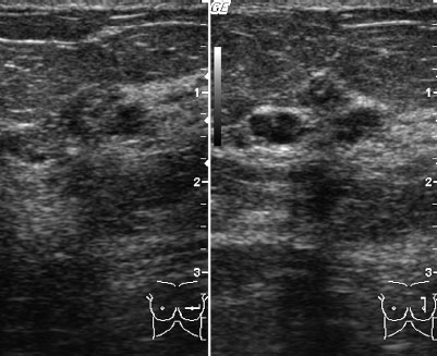
Fig. 65.1 Ultrasound.
Clinical Findings
No findings.
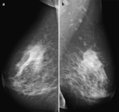
Fig. 65.2 a,b Digital mammography, MLO view [imaging not performed by authors].
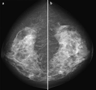
Fig. 65.3 a,b Digital mammography, CC view [imaging not performed by authors].
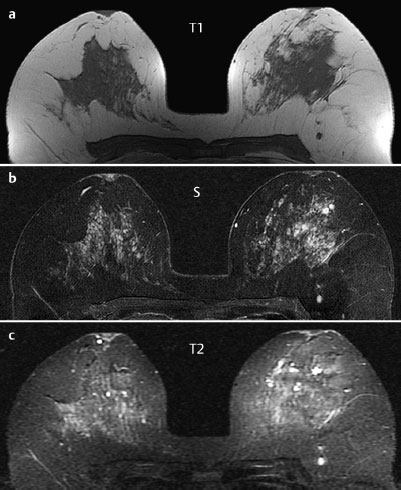
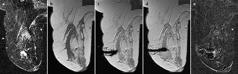
Fig. 65.4 a-c Contrast-enhanced MRI of the breasts.
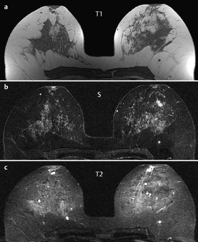
Fig. 65.5 a-c Contrast-enhanced MRI of the breasts.
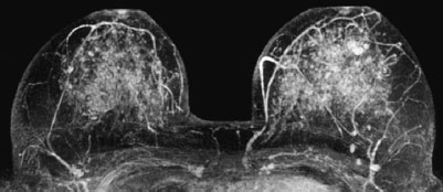
Fig. 65.6 Contrast-enhanced MR mammography. Maximum intensity projection.
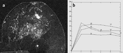
Fig. 65.7 a,b Signal-to-time curves.

|
Please characterize ultrasound, mammography, and MRI findings.
What is your preliminary diagnosis?
What are your next steps? |
This case demonstrates supplementary imaging to investigate suspicious findings in an ultrasound examination performed elsewhere.
Ultrasound
Ultrasound showed a cluster of round, hypoechogenic lesions with linear distal echo shadowing situated in the left breast behind the nipple. US BI-RADS 4.
Mammography
Mammograms showed bilaterally symmetric, inhomogeneously dense parenchyma, ACR type 3. Particularly in the center of the left breast there were no suspicious lesions or densities. Images showed no suspicious microcalcifications and no architectural distortion. BI-RADS right 1/left 1. PGMI: MLO view I (right pectoralis muscle not depicted; inframammary fold not depicted, prominent abdominal skin fold); CC view M (medial parenchyma not fully visible).
MR Mammography
A mass composed of three well-defined, oval, homogeneously enhancing components, each around 1 cm in diameter, was visible in the center of the left breast. It had initial signal increase of 150% and a postinitial washout. In T2-weighted imaging the section of the triple lesion nearest the nipple showed increased signal, while the two sections farther from the nipple had reduced signal. Maximum intensity projection showed a chain of lymph nodes within the breast.
MRI Artifact Category: 1
MRI Density Type: 3
 Differential Diagnosis