Case 74
Indication: Palpable lump in right breast discovered 3 weeks previously.
History: Unremarkable.
Risk profile: No increased risk.
Age: 54 years.
Clinical Findings
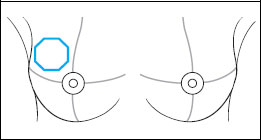
Large, mobile lump in the upper c quadrant of the right breast. No axi lymph nodes palpable.
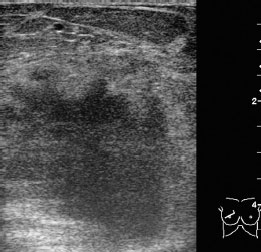
Fig. 74.1 Ultrasound.
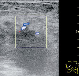
Fig. 74.2 Color-coded Doppler sonography.
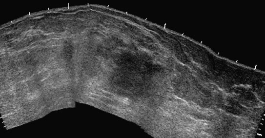
Fig. 74.3 Ultrasound. Panoramic view of the right breast.
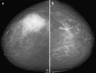
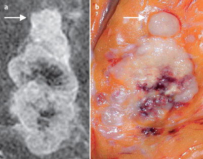
Fig. 74.4a,b Digital mammography, CC view.
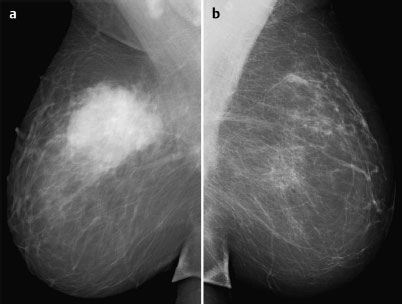
Fig. 74.5a,b Digital mammography, MLO view.
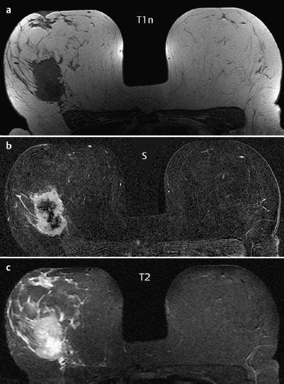
Fig. 74.6a–c Contrast-enhanced MRI of the breasts.
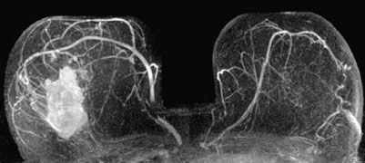
Fig. 74.7 Contrast-enhanced MR mammography. Maximum intensity projection.
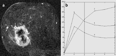
Fig. 74.8a,b Signal-to-time curves.

|
Please characterize ultrasound, mammography, and MRI findings.
What is your preliminary diagnosis?
What are your next steps? |
Clinical evidence already pointed to a definitive diagnosis of breast cancer even before the imaging shown here was carried out. The patient’s statement that the lump was discovered only 3 weeks before the examination was very surprising, since lumps of this size are known to develop over a period of years. However, see for yourself…
Ultrasound
In the area of the palpable mass, ultrasound demonstrated an extensive tumor with multiple criteria of malignancy as well as increased peripheral vascularization in Doppler sonography. The best depiction of the lesion was in panoramic view because of its size. US BI-RADS right 5.
Mammography
The parenchyma was bilaterally predominantly lipomatous, ACR type 1. A massive, ill-defined, hyperdense mass was seen in the right breast. There were no accompanying microcalcifications and no other suspicious findings. BI-RADS right 5/left 1.
MR Mammography (in this case unnecessary)
MRI showed a lobulated, ill-defined tumor in the upper outer quadrant of the right breast, with marked central necrosis. Signal-time curves in the region of interest were typical for malignancy (N.B: The most significant curves in terms of malignancy are always used for the MRM score, even if some individual curves appear normal). Interestingly, in the region of the central necrosis, small hypervascularized nodules were depicted, their development evidently metachronous to the primary tumor growth.
MRI Artifact Category: 1
MRI Density Type: 1
 Preliminary Diagnosis