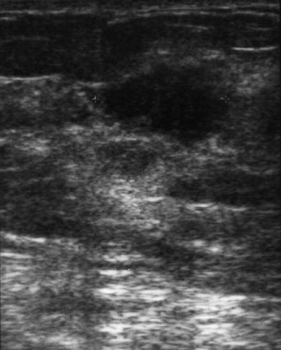
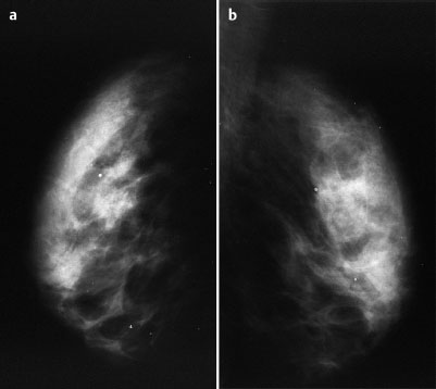
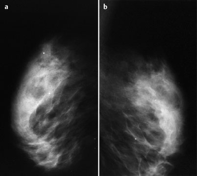
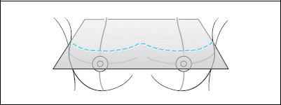
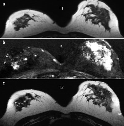
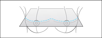
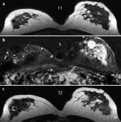
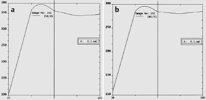
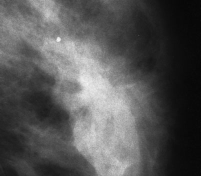
Case 9 Indication: Palpable mass between upper quadrants of the left breast. History: Unremarkable. Risk profile: Breast cancer in grandmother at the age of 82 years. Age: 52 years. Fig. 9.1 Ultrasound image of the palpable mass. Palpable mass of approx. 1.5 cm located in line with the nipple between the upper quadrants of the left breast. Generally lumpy parenchymal texture. Fig. 9.2a,b Conventional mammography, CC view [imaging not performed by authors]. Fig. 9.3a,b Conventional mammography, MLO view [imaging not performed by authors]. Fig. 9.4a–c Contrast-enhanced MRI of the breasts, 3-D technique [imaging not performed by authors]. Fig. 9.5a–c Contrast-enhanced MRI of the breasts, 3-D technique [imaging not performed by authors]. Fig. 9.6a,b Signal-to-time curve [data not prepared by authors]. Please characterize ultrasound, mammography, and MRI findings. What is your preliminary diagnosis? What are your next steps? Corresponding to the palpable resistance there is an irregular hypoechogenic lesion with partially echogenic borders and indeterminate distal echo characteristics in the left breast. US BI-RADS left 4. Mammography showed bilaterally symmetric inhomogeneous dense parenchyma (ACR type 3). There was a hyperdense lesion with a diameter of 1 cm accompanied by architectural distortion in the center of the left breast (Fig. 9.7). Imaging showed no suspicious microcalcifications. BI-RADS right 1/left 4. PGMI: MLO view I (incomplete presentation of the parenchyma; minimal presentation of the pectoral muscle); CC view I (exposure, nipples projected into breast tissue). Corresponding to the clinical and ultrasound findings, MRI demonstrated a round, hypervascularized, partially spiculated lesion 1.3 cm in diameter in the left breast. Initial signal increase of more than 280% (NB: 3-D technique!) and postinitial plateau. There was evident asymmetry of the parenchymal perfusion (left>right). MRI Artifact Category: 2 MRI Density Type: 3* Fig. 9.7 Magnification of the MLO view, left breast.
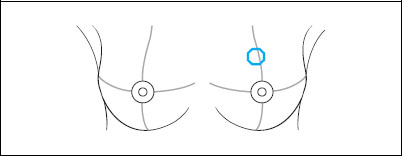
Clinical Findings

Ultrasound
Mammography
MR Mammography
MRM score | Finding | Points |
Shape | round | 1 |
Border | ill-defined | 1 |
CM Distribution | ring (?)* | 2 |
Initial Signal Intensity Increase# | strong | 2 |
Post-initial Signal Intensity Character | plateau | 1 |
MRI score (points) |
| 7 |
MRI BI-RADS |
| 5 |
* Contrast in MR images too high.
# note: 3-D technique
 Preliminary Diagnosis
Breast cancer. Considerably less likely: fibroadenoma.
BI-RADS Categorization | ||
Clinical Findings | right 1 | left 4 |
Ultrasound | right 1 | left 4 |
Mammography | right 1 | left 4 |
MR Mammography | right 1 | left 5 |
BI-RADS Total | right 1 | left 5 |
Procedure recommended elsewhere
(departing from guidelines)
Open biopsy of the left breast for hitrological analysis of the lesion
Procedure according to guidelines
Ultrasound guided core biopsy.
Histology
Fibrocystic mastopathy including an intraductal papilloma (2 mm) and a fibroadenoma (3 mm).
Papilloma and fibroadenoma


Stay updated, free articles. Join our Telegram channel

Full access? Get Clinical Tree


