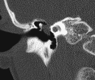
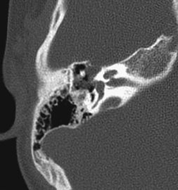
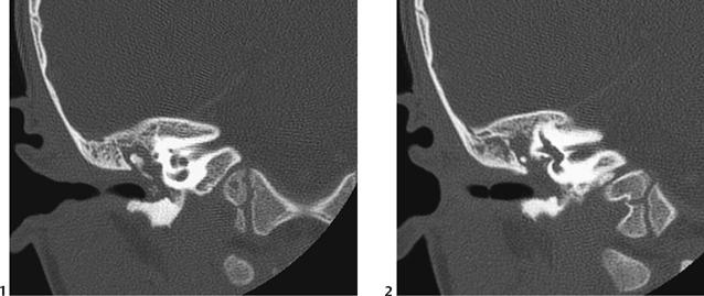
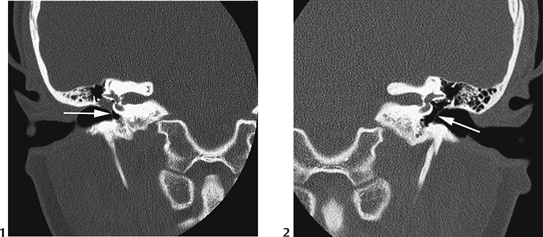
CASE 28 A 9-year-old female presents with a “pearly white” right retrotympanic mass noted during investigation of moderate hearing loss. Figure 28A Figure 28B Noncontrast coronal (Fig. 28A) and axial (Fig. 28B) CT demonstrates a soft tissue mass in the right epi/mesotympanum with erosion of the medial tegmen tympani. Figure 28C Coronal CT (bone algorithm) showing inflammatory soft tissue mass right middle-ear cavity (1). No erosion with soft tissue thickening of the superior external auditory canal (2). Right middle-ear cholesteatoma (Fig. 28C) Congenital cholesteatoma is a keratin-filled sac in the middle ear, located medial to an intact tympanic membrane. Criteria for clinical diagnosis include a white mass medial to a normal tympanic membrane and normal pars tensa and flaccida without prior history of otorrhea, tympanic membrane perforation, or otologic surgery. There is a strong male predominance. Estimated incidence of 0.12 per 100,000. Average age at presentation is between 5 and 7 years. Embryonic rest cell theory proposed by Teed in 1936, who described an epithelial thickening of ectodermal origin in the upper mesotympanum medial to the malleus neck. This epithelial thickening usually involutes and becomes normal endothelium. If this does not occur, then a congenital cholesteatoma may develop. Embryonic epidermal inclusions fail to involute. In theory, they can form in utero, as proven when Michaels in 1986 described a structure in fetal ears at the point of transition between cuboidal and respiratory epithelium and called this “epidermoid” formation. This can be detected normally between 10 and 33 weeks’ gestation; if it persists for more than 33 weeks, is abnormal. Figure 28D Coronal CT showing right middle-ear soft tissue mass (1) with tympanic membrane retraction (arrow). The normal side (2) is shown for comparison. White pearly retrotympanic mass is usually present in the anterior-superior quadrant of the middle ear with an intact tympanic membrane. Presents most commonly with painless otorrhea and hearing loss. Rarely presents with otalgia, headache and fever, vertigo, and facial nerve palsy. Physical findings: white “pearly” retrotympanic mass with an intact tympanic membrane.
Clinical Presentation
Radiologic Findings
Diagnosis
Differential Diagnosis
Discussion
Background
Etiology
Embryology
Clinical Findings
Imaging Findings
Related posts:


Stay updated, free articles. Join our Telegram channel

Full access? Get Clinical Tree


