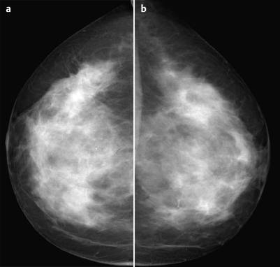
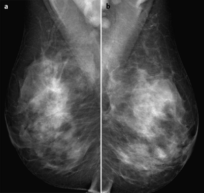
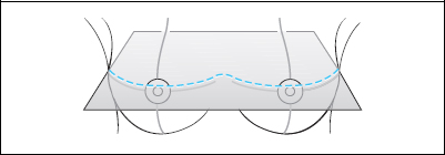
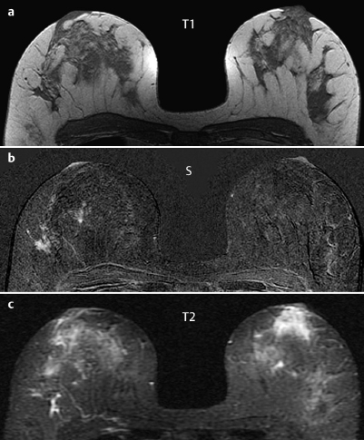
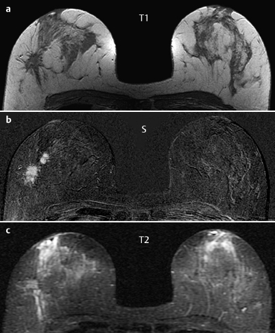
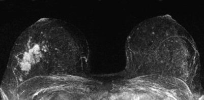
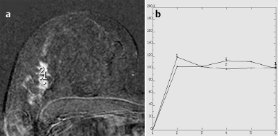
Case 29 Indication: Screening. History: Unremarkable. Risk profile: No increased risk. Age: 54 years. No abnormalities; very firm parenchymal texture in both breasts. Fig. 29.1 a-d Sonography of the right breast. Fig. 29.2a,b Digital mammography, CC view Fig. 29.3a,b Digital mammography, MLO view. Fig. 29.4a-c Contrast-enhanced MRI of the breasts. Fig. 29.5a-c Contrast-enhanced MRI of the breasts. Fig. 29.6 Contrast-enhanced MR mammography. Maximum intensity projection. Fig. 29.7a,b Signal-to-time curves. Please characterize ultrasound, mammography, and MRI findings. What is your preliminary diagnosis? What are your next steps? These are the imaging studies of an asymptomatic woman with no increased risk profile. Ultrasound demonstrated suspect irregular lesions with hyperechoic margins in the lateral region of the right breast. These lesions showed individual distal acoustic shadowing and they disrupted adjoining ligamental structures. There was notable architectural distortion in the inferior region of the right breast depicted in ultrasound. US BI-RADS right 5. Bilaterally extremely dense glandular tissue, ACR type 4. There was an asymmetry of the parenchymal structures with a distortion of the normal architecture and “shrinking sign” in the lateral part of the right breast. Mammography showed no circumscribed mass. No suspicious calcifications. The left breast was normal (BI-RADS right 4/left 1). PGMI: CC view P; MLO view G (inframammary fold incorrectly positioned). In precontrast T1-weighted imaging, MRI reproduced the mammography findings of architectural distortion of the parenchyma and also depicted further nodular formations. Signal in T2-weighted imaging was nonspecific. In the outer quadrants of the right breast, postcontrast imaging depicted partially nodular, partially diffuse multiple enhancing areas with poorly-defined margins and suspect signal behavior. MRI Artifact Category: 2 MRI Density Type: 1
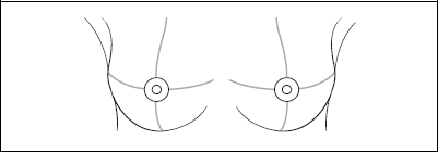
Clinical Findings
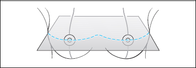

Ultrasound
Mammography
MR Mammography
MRM score | Finding | Points |
Shape | irregular | 1 |
Border | ill-defined | 1 |
CM Distribution | inhomogenous | 1 |
Initial Signal Intensity Increase | strong | 2 |
Post-initial Signal Intensity Character | plateau | 1 |
MRI score (points) |
| 6 |
MRI BI-RADS |
| 5 |
 Preliminary Diagnosis
Extensive carcinoma of the right breast. No differential diagnoses.
Clinical Findings | right 1 | left 1 |
Ultrasound | right 5 | left 1 |
Mammography | right 4 | left 1 |
MR Mammography | right 5 | left 1 |
BI-RADS Total | right 5 | left 1 |
Procedure
Histological evaluation of the suspect findings in the outer quadrants of the right breast, preferably by US-guided percutaneous core biopsy.
Histology of the biopsy specimen (right breast)
Invasive ductal carcinoma.
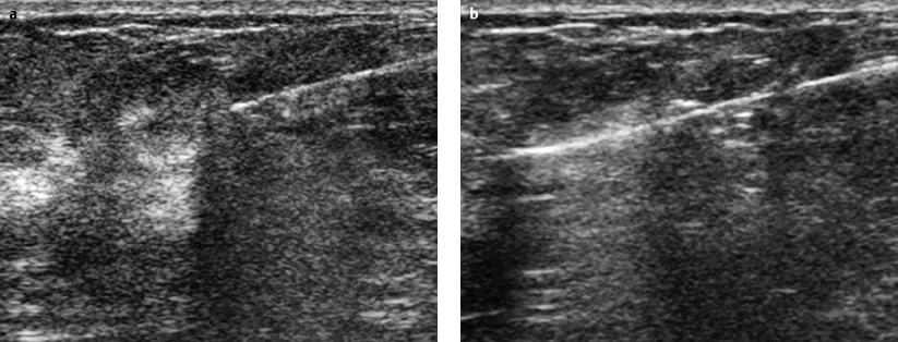
Fig. 29.8a,b US-guided core biopsy. Removal of 5 specimens (14 gauge) using coaxial technique. Pre and post-fire documentation.
Histology
Invasive ductal carcinoma with extensive intraductal component.
IDC pT2 + EIC, pN0, G2.
Stay updated, free articles. Join our Telegram channel

Full access? Get Clinical Tree


