MRM score | Finding | Points |
Shape | round/oval | 0 |
Border | ill-defined | 1 |
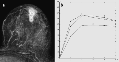
CM Distribution | rim-sign | 2 |
Initial Signal Intensity Increase | strong | 2 |
Post-initial Signal Intensity Character | plateau | 1 |
MRI score (points) |
| 6 |
MRI BI-RADS |
| 5 |
 Preliminary Diagnosis
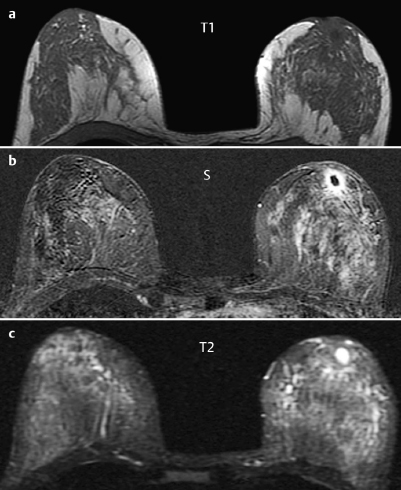
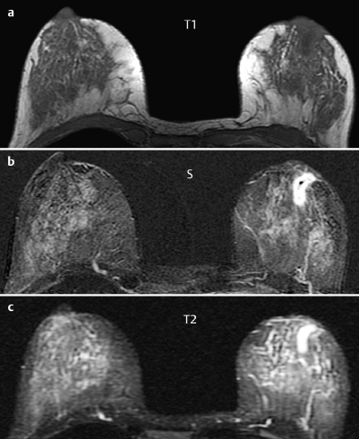
Chronic mastitis with abscesses and blocked milk ducts, duct ectasia.
Differential Diagnosis
Inflammatory carcinoma, Paget disease.
Clinical Findings | right 1 | left 3 |
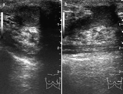
Ultrasound | right 1 | left 3 |
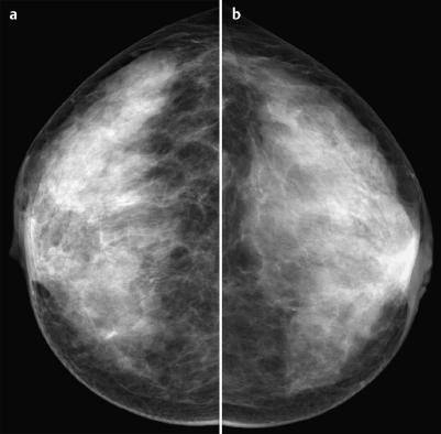
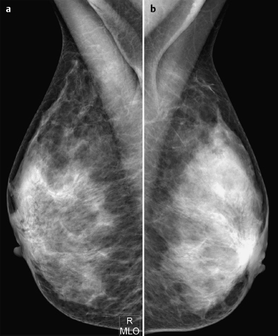
Mammography | right 1 | left 3 |
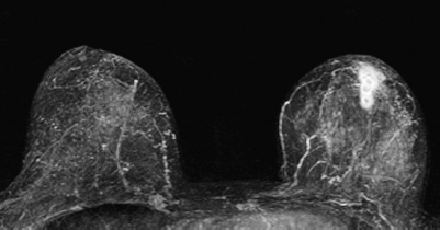
MR Mammography | right 1 | left 5 |
BI-RADS Total | right 1 | left 3 |
Procedure
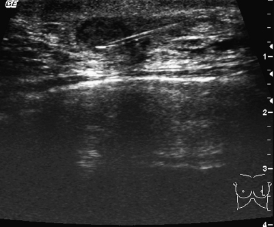
Fine-needle aspiration of the fluid in the region behind the left areola to verify preliminary diagnosis. Bacteriological analysis of the specimen to identify the pathogen and determine possible resistance to antibiotic therapy.
Cytology
Infected inspissated cyst.
Fig. 30.8 US-guided FNAP of the abscess.
Diagnosis
Empyemic abscess in milk duct.
Stay updated, free articles. Join our Telegram channel

Full access? Get Clinical Tree


