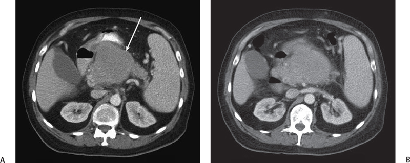
CASE 30 A 75-year-old man with a history of multiple myeloma presents with abdominal pain. Fig. 30.1 (A,B) Axial contrast-enhanced CT image of the abdomen shows a round, well-defined mass in the pancreatic head and body (arrow) extending to the third portion of the duodenum. The splenic vein is encased by the pancreatic mass and can be appreciated only in its proximal part. Contrast-enhanced computed tomography (CT) images of the pancreas (Fig. 30.1) show a large mass centered in the pancreas and causing extrinsic compression of the duodenum; the splenic vein appears to be obliterated in the region of the mass. The tail of the pancreas is spared and shows no atrophy or pancreatic duct dilatation. Extramedullary plasmacytoma Pancreatic mass:
Clinical Presentation
Radiologic Findings
Diagnosis
Differential Diagnosis
Discussion
Background
Related posts:


Stay updated, free articles. Join our Telegram channel

Full access? Get Clinical Tree


