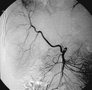
CASE 30 A 45-year-old male presented to interventional radiology for chemoem-bolization to treat hepatocellular carcinoma involving the right lobe of the liver. Figure 30-1 Selected arteriogram shows replaced right hepatic artery originating from the superior mesenteric artery and perfusing a hepatoma. The right common femoral artery was punctured using the Seldinger technique, and a 5-French (F) sheath was inserted. The celiac artery was catheterized using an RC-1 (Boston Scientific, Natick, Massachusetts), and angiography was performed showing absence of flow to the right lobe of the liver. Subsequently, the superior mesenteric artery (SMA) was catheterized, and angiography showed a replaced right hepatic artery originating from the SMA and supplying the tumor (Fig. 30-1). Replaced right hepatic artery originating from the SMA feeding hepatocellular carcinoma. Using a 3F microcatheter, the subsegmental arteries supplying the hepatocellular carcinoma were superselected. Chemoembolization was performed by first injecting a mixture of doxorubicin and Ethiodol, followed by cisplatin and mitomycin.
Clinical Presentation
Radiologic Studies
Conventional Angiography
Diagnosis
Treatment
| Variant | Incidence |
| Right HA from SMA (Fig. 30-1) | < 16% |
| Left HA from left gastric A (Fig. 30-2) | < 10% |