30 Ventriculus Terminalis
30.1 Case Presentation
An 18-month-old female patient presents with small shallow sacral dimple.
30.2 Imaging Analysis
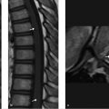
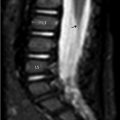
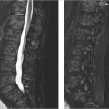
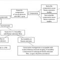
MRI of an 18-month-old female patient with sagittal T1-weighted (T1w; ▶ Fig. 30.1a), sagittal short tau inversion recovery (STIR; ▶ Fig. 30.1b), axial T1w (▶ Fig. 30.1c), and axial T2-weighted (T2w; ▶ Fig. 30.1d) images. This image shows a linear-shaped cystic structure within the central portion of the conus medullaris that has similar signal intensity to cerebrospinal fluid (CSF) on T1w and T2w images (arrowheads). This structure corresponds to an expanded central canal without any spinal cord signal abnormality. It is important to note that conus medullaris is in normal position and the filum terminale is normal in appearance (arrows).

30.3 Differential Diagnosis
Ventriculus terminalis:
Normal variant representing a focal dilatation of the central canal in the conus.
Transient dilatation of the central canal:
Normal variant in which there is a slight transient dilatation of the central canal during the first week of life. 1 , 2
Cystic spinal cord neoplasm:
The imaging landmark is cord signal abnormality, mass effect, neurologic symptoms, and contrast enhancement in solid portions.
Syringohydromyelia:
Cystic dilatation of the central canal that could be isolated or associated with congenital anomalies in up to 30% of cases. 3
Myelomalacia:
Cord atrophy secondary to previous vascular, traumatic, or other injury.
Related posts:
Stay updated, free articles. Join our Telegram channel

Full access? Get Clinical Tree


