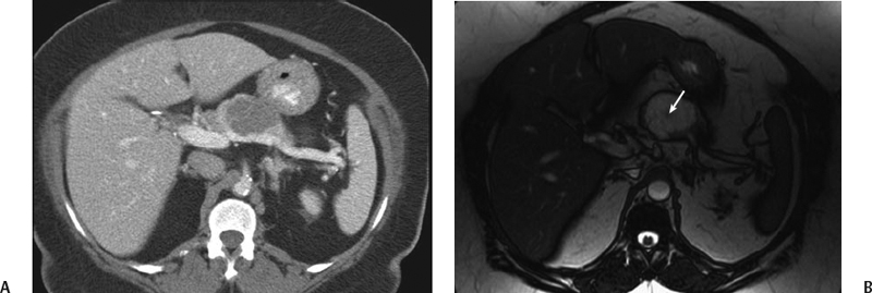
CASE 31 A 63-year-old woman complains of epigastric discomfort. Fig. 31.1 (A) Axial postcontrast CT image shows a well-defined low-density lesion with fine septations in the body of the pancreas. (B) Axial T2-weighted image in the same patient shows a well-defined hyperintense mass lesion within the body of the pancreas with a central scar (arrow). Postcontrast axial computed tomography (CT) and magnetic resonance (MR) (Fig. 31.1) images of the pancreas show a well-defined cystic lesion in the body of the pancreas with enhancing thin septation and a central scar. Microcystic adenoma (serous cystadenoma) of the pancreas
Clinical Presentation
Radiologic Findings
Diagnosis
Differential Diagnosis
Discussion
Background
Related posts:


Stay updated, free articles. Join our Telegram channel

Full access? Get Clinical Tree


