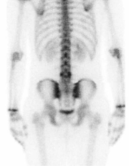
CASE 31 A 17-year-old male adolescent presents with a history of low back pain of 5 months’ duration. A bone scan is requested (Figs. 31.1, 31.2, and 31.3). Fig. 31.1 • A 20 mCi dose of 99mTc-MDP is administered intravenously. • Whole-body images of the skeleton are obtained 3 hours after tracer administration. • A 1024 × 256 matrix is used for whole-body images. • Emphasize the importance of oral hydration to improve soft tissue and bladder clearance. • SPECT of the lumbar spine is obtained 4 hours after tracer injection: 64 stops, 25 seconds per stop, and a 360-degree rotation.
Clinical Presentation
Technique
Related posts:


Stay updated, free articles. Join our Telegram channel

Full access? Get Clinical Tree


