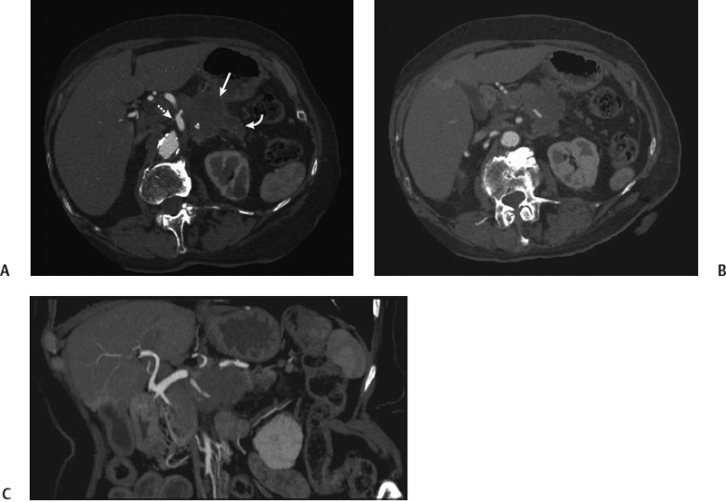
CASE 32 A 75-year-old man presents with mild abdominal pain, loss of appetite, and weight loss in the last few months. Fig. 32.1 (A–C) Axial contrast-enhanced CT images show a large, enhancing mass (arrow) within the body of the pancreas. The distal pancreatic parenchyma appears markedly atrophic with a dilated main pancreatic duct (curved arrow). The mass encases a portion of the celiac artery (dotted arrow) but spares the mesenteric artery. No calcifications are noted. There is no local lymphoadenopathy. Axial contrast-enhanced computed tomography (CT) images (Fig. 32.1) show a hypodense, peripherally enhancing mass within the body of the pancreas associated with marked pancreatic ductal dilatation and atrophy of the pancreas proximal to the mass. Massive peripancreatic vessel encasement is seen. Ductal adenocarcinoma of the pancreatic head Ductal pancreatic adenocarcinomas are the fifth leading cause of death in Western countries, representing almost 75 to 85% of nonendocrine pancreatic malignancies. These tumors, more frequently located in the head of the pancreas (60–70% of cases), usually affect elderly men in their 7th or 8th decade.
Clinical Presentation
Radiologic Findings
Diagnosis
Differential Diagnosis
Discussion
Background
Clinical Findings
Related posts:


Stay updated, free articles. Join our Telegram channel

Full access? Get Clinical Tree


