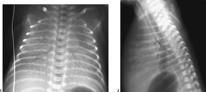
CASE 32 At the second day of life, a 27-week-gestational-age premature infant presents with progressive respiratory distress. Figure 32A Frontal radiographic view of the intubated premature infant demonstrates diffuse, hazy “ground-glass,” symmetric pulmonary opacification, and air space disease (Fig. 32A1). The lung volume is small due to diffuse microatelectasis from surfactant deficiency. Distended and prominent central “air bronchograms” are seen due to the relatively high-pressure ventilation and peripheral poor alveolar compliance (Fig. 32A2). Hyaline membrane disease (HMD) HMD occurs more commonly in low-birth-weight (<2500 g) premature infants born earlier than 38 weeks of gestation. HMD is caused by a combination of factors. The premature lung has a paucity of type 2 pneumocytes, which form and secrete surfactant. Compared with a mature infant, the lung of a premature infant has thicker interstitium, which does not permit efficient gas exchange. This results in increased work of breathing and an increased metabolic rate. While trying to maintain a very high metabolic rate for growth, the premature infant has little respiratory capacity and poor energy reserves. These features combine to result in early respiratory failure. Furthermore, conventional treatment has used high-pressure ventilation and high oxygen concentrations, both of which cause direct lung damage. Part of this injury process involves an inflammatory reaction, including the formation of proteinaceous exudates, which fill the alveoli and constitute the “hyaline membranes.”
Clinical Presentation

Radiologic Findings
Diagnosis
Differential Diagnosis
Discussion
Background
Etiology and Pathophysiology
Imaging Findings
RADIOGRAPHY
Stay updated, free articles. Join our Telegram channel

Full access? Get Clinical Tree


