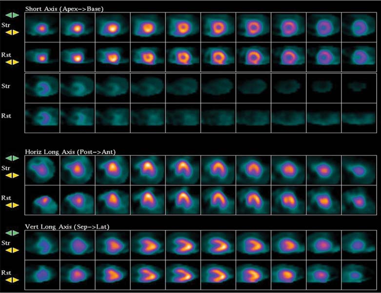
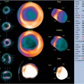
CASE 32 An 85-year-old woman with no known coronary artery disease (CAD) is referred for an exercise myocardial perfusion single-photon emission tomography (SPECT) study to evaluate for nonanginal chest pain. Her cardiac risk factors include hypertension. The resting electrocardiogram (ECG) shows normal sinus rhythm and nonspecific T-wave abnormalities. She is on lisinopril and hydrochlorothiazide at the time of testing. Fig. 32.1 • The patient had nothing to eat within 4 hours of the test. • Rest images were acquired 40 minutes after the intravenous injection of 10 mCi of 99mTc-sestamibi. Images were acquired in the supine position with a two-headed gamma camera with step-and-shoot rotation, 32 projections over a 90-degree arc for each head (64 projections over a 180-degree arc), 30 seconds per projection, and a 64 × 64 matrix. • Exercise: 9 minutes, 0 second on a Bruce protocol (10.1 METs [metabolic workloads]). • A 33 mCi dose of 99mTc-sestamibi was injected during peak stress. • At 45 minutes after the exercise injection of radiotracer, image data were acquired with settings similar to those used for rest imaging. Gated images were acquired at 8 frames per cardiac cycle. • Transverse images were reconstructed with a Butterworth filter (order of 5 and cutoff frequency of 0.792 cycles per pixel) for the rest and stress studies. The heart rate increased from 59 beats/min at rest to a peak of 116 beats/min (86% of the age-predicted maximal heart rate), and the blood pressure increased from 140/60 mm Hg at rest to 160/80 mm Hg at peak exercise (rate-pressure product of 18,560). Exercise was terminated because of fatigue. Baseline ECG was normal. There were no symptoms of ischemia. The perfusion images are shown in Fig. 32.1.
Clinical Presentation
Technique
Image Interpretation
![]()
Stay updated, free articles. Join our Telegram channel

Full access? Get Clinical Tree


