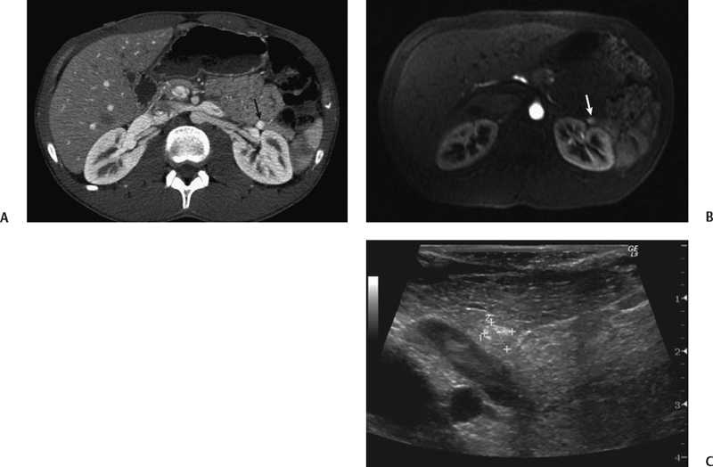
CASE 33 A 53-year-old woman presents with mild abdominal pain, diaphoresis, and palpitations. Fig. 33.1 (A) Axial contrast-enhanced CT image shows a small arterially enhancing lesion in the tail of the pancreas (arrow). (B) The lesion is also seen on an early arterial phase gadolinium-enhanced MRI. (C) The patient underwent intraoperative ultrasound prior to resection, which confirmed the lesion. Axial contrast-enhanced computed tomography (CT) images (Fig. 33.1) show a small arterially enhancing lesion in the tail of the pancreas. The lesion is also seen on an early arterial-phase gadolinium-enhanced magnetic resonance (MR) image. Intraoperative ultrasound confirmed the lesion prior to resection. Islet cell tumor of the pancreas (insulinoma)
Clinical Presentation
Radiologic Findings
Diagnosis
Differential Diagnosis
Discussion
Background
Related posts:


Stay updated, free articles. Join our Telegram channel

Full access? Get Clinical Tree


