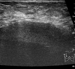
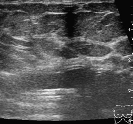
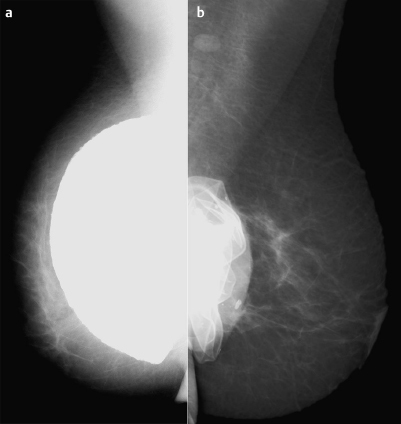
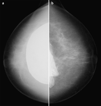
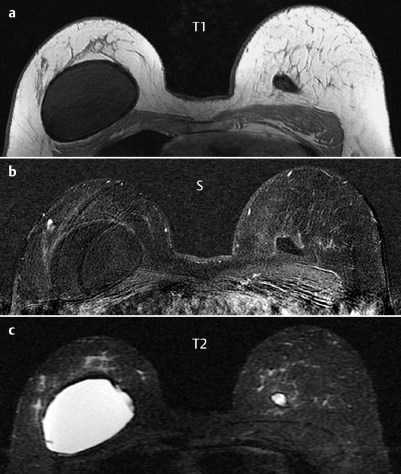
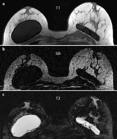
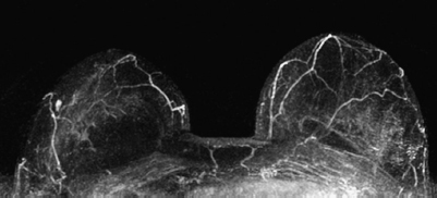
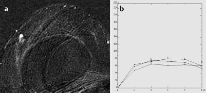
Case 34 Indication: Screening. History: Bilateral implants 30 years previously to correct anisomastia. Risk profile: Not increased. Age: 53 years. Regular asymptomatic scars bilaterally. Fig. 34.1 Ultrasound image, right breast. Fig. 34.2 Ultrasound image, left breast. Fig. 34.3a,b Digital mammograpy, MLO view. Fig. 34.4a,b Digital mammography, CC view. Fig. 34.5a–c Contrast-enhanced MRI of the breasts. Fig. 34.6a–c Contrast-enhanced MRI of the breasts. Fig. 34.7 Contrast-enhanced MR mammography. Maximum intensity projection. Fig. 34.8a,b Signal-to-time curves. Please characterize ultrasound, mammography, and MRI findings. What kind of implants do you see? What is your preliminary diagnosis? What are your next steps? Ultrasound showed that the left prosthesis had a markedly smaller volume than the right. There were no other unusual findings. USBI-RADS right 1/left 1. Images showed a fibroglandular parenchyma, ACR type 2. There were no suspicious lesions and no architectural distortions. Mammography demonstrated no suspicious microcalcifications. Left mammography showed a collapsed prosthesis with clumped calcifications. BI-RADS right 1/left 1. PGMI is not defined for implantimages. MRI documented the collapsed left implant. It depicted a single-lumen saline-filled implant (no adequate signal in silicon sequences, Fig. 34.6b). MRI also depicted a well-defined, hypervascularized focus of 1 cm diameter in the upper outer quadrant of the right breast, which showed nonspecific signal behavior after contrast administration. T2 signal in the focal area was increased. This focus had a lipomatous central area in precontrast Tl -weighted imaging. Have you been able to determine the type of prosthesis involved here? MRI Artifact Category: 2 MRI Density Type: 1
Clinical Findings

Ultrasound
Mammography
MR Mammography
MRM score | Finding right | Points |
Shape | round | 0 |
Border | well-defined | 0 |
CM Distribution | homogenous | 0 |
Initial Signal Intensity Increase | moderate | 1 |
Post-initial Signal Intensity Character | plateau | 1 |
MRI score (points) |
| 2 |
MRI BI-RADS |
| 2 |
 Diagnosis
Left breast: Complete rupture of the implant (envelope and capsule). No differential diagnosis.
Right breast: Nonspecific lymphadenitis. No differential diagnosis.
Clinical Findings | right 1 | left 1 |
Ultrasound | right 1 | left 1 |
Mammography | right 1 | left 1 |
MR Mammography | right 2 | left 1 |
BI-RADS Total | right 2 | left 1 |
Procedure
None. This woman had saline-only prostheses in both breasts. No intervention was necessary following the complete rupture of the left implant, since the saline content was completely resorbed. The hypervascularized focus in the right breast could be clearly identified as a lymph node due to the fatty hilum visible at its center in MRI. This is therefore categorized as a harmless ancillary finding.
Remark
Luckily the rupture occurred in the larger breast. Aesthetically, no intervention was required. The uneven calcifications within the capsule did not show criteria of malignancy.
Diagnosis (without operative or histopathological verification)
Left: Complete rupture of saline implant.
Right: Lymphadenitis.
Stay updated, free articles. Join our Telegram channel

Full access? Get Clinical Tree


