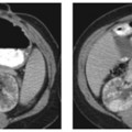
CASE 34 A 70-year-old man presents with epigastric pain 1 year after left nephrectomy for renal cell carcinoma. Fig. 34.1 Axial contrast-enhanced image in the early arterial phase of enhancement in this patient with left nephrectomy shows a well-defined enhancing hypervascular lesion in the body of the pancreas (arrow). A well-defined homogeneously enhancing hypervascular lesion is seen in the body of the pancreas anteriorly. Diffuse fatty atrophy of the pancreas is present. Note the surgical clips in the left renal fossa at the location of the left renal vascular pedicle consistent with a past history of nephrectomy for renal cell carcinoma (Fig. 34.1). Metastatic renal cell carcinoma to the pancreas
Clinical Presentation

Radiologic Findings
Diagnosis
Differential Diagnosis
Discussion
Background
Stay updated, free articles. Join our Telegram channel

Full access? Get Clinical Tree


