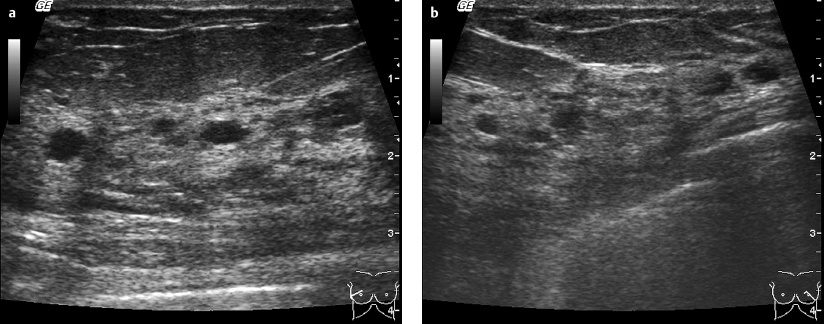
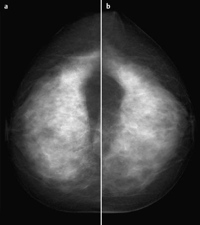
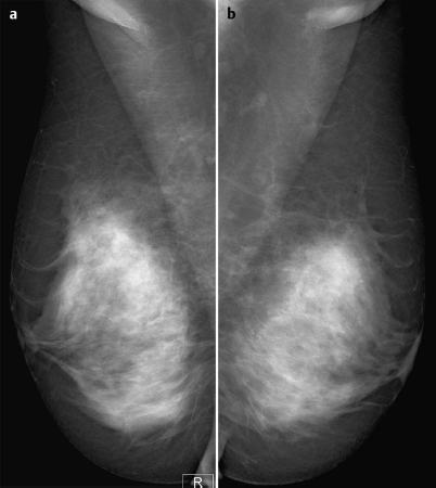
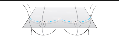
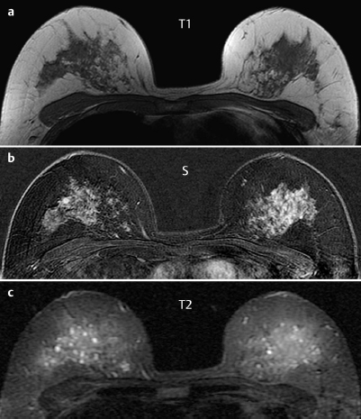
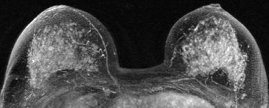
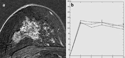
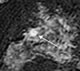
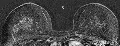
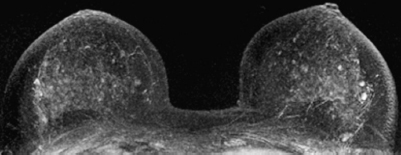
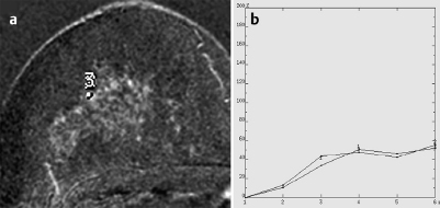
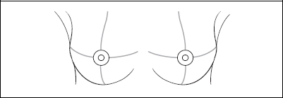
Case 35 Indication: Screening. History: Unremarkable; currently undergoing hormone replacement therapy. Risk profile: Breast cancer in an aunt and two cousins. Age: 53 years. Normal. Fig. 35.1a,b Sonography. Fig. 35.2 Digital mammography, CC view. Fig. 35.3 Digital mammography, MLO view. Fig. 35.4a–c Contrast-enhanced MRI of the breasts. Fig. 35.5 Contrast-enhanced MR mammography. Maximum intensity projection. Fig. 35.6a,b Signal-to-time curves. Please characterize the ultrasound, mammography and MRI findings. What is your preliminary diagnosis? What are your next steps? Ultrasound showed several cysts in the upper outer quadrants of both breasts. There were no suspicious findings. US BI-RADS right 2/left 2. Mammograms showed bilaterally symmetric, extremely dense tissue, ACR type 4. Under the limitations imposed by these conditions, no abnormalities were visible (no density, no mass, no microcalcifications, no architectural distortion). BI-RADS right 1/leftl.PGMI: CC view I (incomplete presentation of the left inner quadrant); MLO view G (inframammary fold and left nipple incorrectly positioned). Both breasts showed generally strong enhancement of the glandular tissue. In the upper outer quadrant of the right breast there was a circumscribed enhancing lesion of 5 mm diameter with multiple criteria of malignancy (Fig. 35.7). MRI Artifact Category: 2 MRI Density Type: 4 Fig. 35.7 Lesion in the upper outer quadrant of the right breast.
Clinical Findings

Ultrasound
Mammography
MR Mammography
MRM score | Finding | Points |
Shape | round | 0 |
Border | well-defined | 1 |
CM Distribution | rimsign | 0 |
Initial Signal Intensity Increase | strong | 2 |
Post-initial Signal Intensity Character | wash-out | 2 |
MRI score (points) |
| 7 |
MRI BI-RADS |
| 5 |
 Differential Diagnosis
Adenosis (hormonally induced?), carcinoma.
Clinical Findings | right 1 | left 1 |
Ultrasound | right 2 | left 2 |
Mammography | right 1 | left 1 |
MR Mammography | right 5 | left 1 |
BI-RADS Total | right 5 | left 1 |
Procedure (Option 1)
MR-guided percutaneous vacuum biopsy of the focal enhancing lesion in the upper outer quadrant of the right breast.
Alternative (Option 2)
Repeat imaging studies 4-6 weeks after stopping the hormone replacement therapy (Figs. 35.8–35.10).
Fig. 35.8 MR imaging six weeks after cease of hormone replacement
Fig. 35.9 Maximum intensity projection after cease of hormone retherapy, placement therapy.
Fig. 35.10a,b Signal-to-time curves after cease of hormone replacement therapy.
Diagnosis (without histopathological verification)
Hormonally-induced focal enhancement in the right breast. Normalization after stopping hormone replacement therapy.
Stay updated, free articles. Join our Telegram channel

Full access? Get Clinical Tree


