CASE 35
Clinical Presentation
A 58-year-old man presents with a history of coronary artery disease and prior percutaneous coronary intervention and stent placement in the right coronary artery. He has no chest pain or shortness of breath. Medications are aspirin, clopidogrel, β-blockers, and calcium channel blockers.
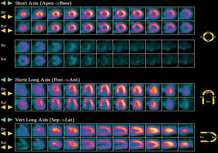
Fig. 35.1
Technique
• The patient had nothing to eat within 4 hours of the test.
• Rest images were acquired 40 minutes after the intravenous injection of 10 mCi of 99mTc-sestamibi. Images were acquired in the supine position with a two-headed gamma camera with step-and-shoot rotation, 32 projections over a 90-degree arc for each head (64 projections over a 180-degree arc), 30 seconds per projection, and a 64 × 64 matrix.
• Exercise: 9 minutes, 4 seconds on a Bruce protocol (12.8 METs [metabolic workloads]).
• Heart rate, blood pressure, and 12-lead ECG were recorded at baseline and every minute thereafter during stress.