CASE 35 A 70-year-old man presents with abdominal pain and weight loss in the last few months. Fig. 35.1 (A) An axial contrast-enhanced CT image of the abdomen demonstrates a large, encapsulated, rounded mass localized in the head and neck of the pancreas. The mass is extremely heterogeneous, as it contains both areas of fluid and soft tissue attenuation; a thin capsule is observed and presents mild enhancement as well as the peripheral solid intratumoral components. The pancreatic parenchyma is otherwise unremarkable, and no sign of pancreatic duct dilatation is documented. (B) Axial image through the liver in the same patient shows a well-demarcated lesion in the left hepatic lobe of the liver with attenuation characteristics similar to the primary pancreatic lesion. This was found to represent a solitary metastasis on surgical biopsy. Axial contrast-enhanced computed tomography (CT) images of the upper abdomen (Fig. 35.1) show a large, well-defined, round pancreatic mass centered within the head and neck; the lesion has areas of low and high attenuation. A similar appearing focal lesion is also seen in the liver. Solid papillary epithelial neoplasm (SPEN)
Clinical Presentation
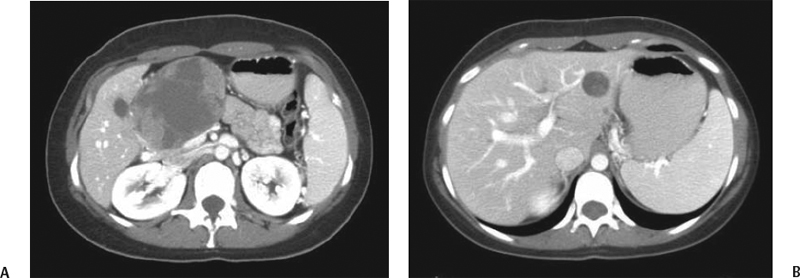
Radiologic Findings
Diagnosis
Differential Diagnosis
Discussion
Background
Related posts:


Stay updated, free articles. Join our Telegram channel

Full access? Get Clinical Tree


