CASE 36
Clinical Presentation
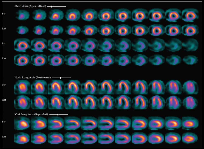
A 68-year-old man is referred for the evaluation of atypical chest pain. The patient denies shortness of breath. His past medical history includes hypertension, atrial fibrillation, and diabetes. He is on aspirin, insulin, metoprolol, and a calcium channel blocker.

Fig. 36.1
Technique
• The patient had nothing to eat within 4 hours of the test. The β-blocker (metoprolol) was withheld on the day of the test. Caffeinated beverages were withheld for 24 hours before the test.
• Rest images were acquired 40 minutes after the intravenous injection of 10 mCi of 99mTc-sestamibi. Images were acquired in the supine position with a two-headed gamma camera with step-and-shoot rotation, 32 projections over a 90-degree arc for each head (64 projections over a 180-degree arc), 30 seconds per projection, and a 64 × 64 matrix.
• A 6-minute adenosine infusion was done.
• Heart rate, blood pressure, and 12-lead ECG were recorded at baseline and every minute thereafter during stress.
• A 28 mCi dose of 99mTc-sestamibi was injected during peak stress (at minute 3).
• Images were acquired 45 minutes after tracer injection. Acquisition protocols for post-stress supine imaging used 32 stops at 25 seconds per stop and a dual-head camera.
• The study was not gated because of the irregular heart rate caused by atrial fibrillation.
• CT images were obtained with a SPECT/CT system. A low-dose nongated CT scan of the chest was obtained for attenuation correction (AC) with the following parameters: scan length, 15 cm; rotation time, 0.5 second; total scan time, 3.9 seconds; tube voltage, 140 kV; and slice thickness, 5 mm. Attenuation correction was performed with the standard reconstruction software.
Image Interpretation


