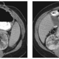
CASE 36 A 58-year-old Caucasian woman presented with a history of epigastric discomfort, anorexia, and weight loss over 3 to 4 months and recent-onset jaundice. Fig. 36.1 Axial contrast-enhanced CT image of the pancreas shows a well-defined fluid density, multicystic mass lesion in the body and tail of the pancreas with thick enhancing nodular wall, septum, and mural excrescences. An axial contrast-enhanced computed tomography (CT) image of the pancreas shows a well-defined fluid density, multicystic lesion in the body and tail of the pancreas with thick, enhancing nodular wall, septum, and mural excrescences (Fig. 36.1). Pancreatic mucinous cystadenocarcinoma Mucinous cystadenocarcinomas are the most common cystic neoplasms seen in the pancreas and account for 50% of all pancreatic cystic neoplasms.
Clinical Presentation

Radiologic Findings
Diagnosis
Differential Diagnosis
Discussion
Background
Clinical Findings
Stay updated, free articles. Join our Telegram channel

Full access? Get Clinical Tree


