36 Renal Cyst


10.1055/b-0040-176872

36 Renal Cyst

Steven S. Chua, Chakradhar R. Thupili, Kaustubh G. Shiralkar, and Eduardo J. Matta

36.1 Case Presentation


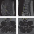
A 71-year-old man presents with lower back pain. An MRI of the lumbar spine without contrast was performed to evaluate for radiculopathy (▶ Fig. 36.1; also see ▶ Table 36.1).

Fig. 36.1 T1-weighted (a) and T2-weighted (b) axial images of the lumbar spine demonstrate renal cystic lesions at the level of the mid to inferior pole of the left kidney. A slightly T1 intermediate to hyperintense left inferomedial 2-cm renal cyst (white arrow in a) is T2 hypointense (white arrow in b) in the left kidney. A T1 hypointense left inferoposterior renal cyst partially seen (yellow arrow in a) is T2 hyperintense (yellow arrow in b).


Table 36.1 Differential diagnosis for cystic lesions of the kidney 1

Differential diagnosis


Comments


Cyst


This structure is the most common lesion of the kidney. Cysts can range from benign to complex, classified by the Bosniak criteria, 2 , 3 , 4 by CT or MR. Benign cysts can be simple with fluid, contain varying amounts of proteinaceous or hemorrhage components, to minimally complex with septations and thin calcifications, to complex with thickened irregular septations with enhancing nodularities to cystic renal cell carcinoma


Renal sinus cyst


This term refers to cysts found in the renal sinus and include parapelvic cyst (cyst extending from the renal parenchyma into the renal hilum) vs. peripelvic cyst (lymphatic cysts extending from the hilum into the renal cortex), and can mimic hydronephrosis. An excretory phase with contrast in the renal collecting systems can show that hydronephrosis is not present with these cysts not opacifying with contrast as they are not connected to the collecting system


Calyceal diverticulum


A calyceal diverticulum extends from and is connected to the renal collecting system and will opacify with contrast in the excretory phase of a contrast-enhanced CT/MR examination. Frequently, milk of calcium or calcium deposits can be found in this herniated sac


Mass


This term usually refers to a solid lesion in the kidney and can range from primary renal cell carcinoma (RCC), urothelial cancer, lymphoma, and inflammatory myofibroblastic tumors to metastatic disease. Tumors that metastasize to the kidney include melanoma and solid tumors of the breast, lung, and gastrointestinal and genitourinary systems. More benign-appearing lesions of the kidney include angiomyolipomas and oncocytomas. Angiomyolipomas show macroscopic fat, which will demonstrate loss of signal in fat-saturated sequences and may also show microscopic fat, which will demonstrate loss of signal in the in- and opposed-phase sequences, and generally do not contain calcifications, unlike fat-containing RCCs, which can contain calcification. Oncocytomas may show a stellate scar and enhance avidly


Pseudotumor


Pseudotumors include hypertrophied columns of Bertin, an extension of the cortex into the medulla of the kidney, which enhance similar to renal parenchyma on all phases. In addition, the dromedary bulging from the interpolar region of the left kidney and fetal lobulations are also examples. No further follow-up are required


Focal infection


Focal pyelonephritis or intrarenal abscesses are also considerations for renal masses. Typically, if pyelonephritis or intrarenal abscesses were discovered on imaging, a follow-up scan after treatment with a CT abdomen and pelvis with contrast is performed


Pseudoaneurysm


Pseudoaneurysm of the renal artery or renal vein can have masslike appearance in the kidney especially in noncontrast images. However, on contrast imaging, these lesions follow the enhancement profile of the vessels


36.2 Diagnostic Testing Needed


When renal cysts are identified in lumbar spine MRI without contrast, they may be further evaluated with renal ultrasound, or renal mass protocol CT/MR, depending on their complexity and size. Ideally, if prior imaging is available, comparison can be helpful to determine stability versus changes. Fluid density cysts that are T2 hyperintense and T1 hypointense are most certainly benign and require no further follow-up especially if these are less than 1 cm, and follow fluid signal like cerebrospinal fluid (CSF) on T2 and T1 sequences, 5 have no solid components, and have well-defined borders. Fluid density cysts greater than 1 cm or those lesions that are partially imaged can be evaluated by renal ultrasound. Lesions without fluid signal or lesions on MRI that are greater than 1 cm can be evaluated by CT/MR renal mass protocol. 5 , 6


On nonenhanced CT, homogenous lesions with internal attenuation less than 20 HU (simple cyst) or greater than 70 HU (hemorrhagic cysts) are considered benign and require no further follow-up, but if the lesions have internal attenuation of between 20 and 70 HU, they are deemed indeterminate and require further workup. 7 , 8 Studies have shown that homogeneous renal lesions greater than 70 HU on nonenhanced CT have a greater than 99.9% of being a hemorrhagic cyst than renal cell carcinoma. 9 This is pertinent as renal cysts can be seen on noncontrast CT of the lumbar spine.


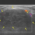
Because the patient had a history of treated lung cancer, a PET/CT was performed to evaluate for the presence of residual disease and a CT abdomen and pelvis with contrast was performed for abdominal pain. Typically, PET/CT examinations are not performed to evaluate renal cysts, but in our patient’s case, the PET/CT examination was useful because it allowed for comparison of the renal cysts. Therefore, the left renal cysts seen on this MRI study can then be compared to these studies.


Given that the left inferomedial cyst was hyperdense in the noncontrast image (white arrow in ▶ Fig. 36.2a) and was T1 hyperintense and T2 hypointense, this structure is most likely a proteinaceous/hemorrhagic cyst and requires no further follow-up. Similarly, the T2 hyperintense and T1 hypointense nonenhancing fluid density inferoposterior cyst is a simple cyst, and requires no further follow-up.

Fig. 36.2 A previous PET/CT fusion axial image of the abdomen (a) at the same level as the prior lumbar spine MRI and a recent contrast-enhanced CT image of the abdomen (b) demonstrate the presence of a hyperdense cyst in the left kidney (white arrow in a and b), corresponding to the previous T1 hyperintense and T2 hypointense cyst (white arrow in ▶ Fig. 36.1a,b) and a hypodense cyst (yellow arrow in a and b) corresponding to the previous T1 hypointense and T2 hyperintense cyst (yellow arrow in ▶ Fig. 36.1a,b) in the left kidney. Image (b) also demonstrates a few fluid density nonenhancing cysts (marked by yellow asterisks) in the bilateral kidneys. None of the left renal cysts demonstrate fluorodeoxyglucose avidity.

Only gold members can continue reading. Log In or Register to continue

Stay updated, free articles. Join our Telegram channel

Jun 28, 2020 | Posted by in NEUROLOGICAL IMAGING | Comments Off on 36 Renal Cyst

Full access? Get Clinical Tree

Get Clinical Tree app for offline access